
Basic Sciences
Electron Microscopy
 Officer-in-Charge: Dr.Sorab N. Dalal
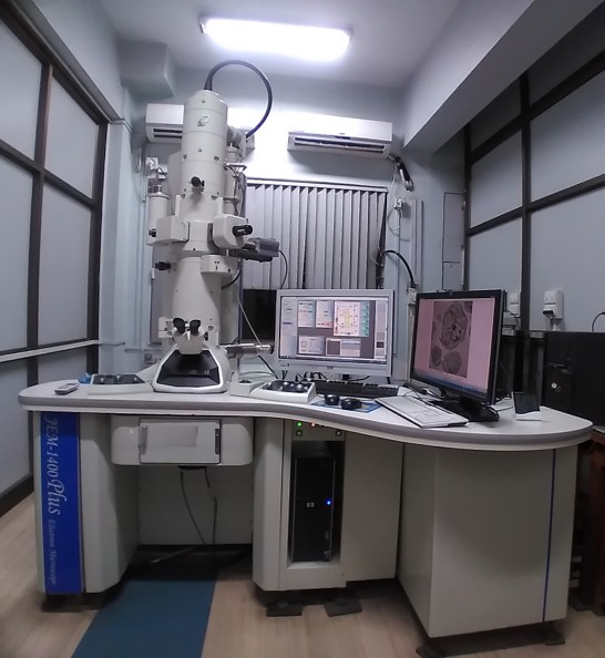
The Electron Microscope Facility at ACTREC is equipped with JEM 1400 PLUS, a Transmission electron microscope (JEOL, Japan) functioning at 120kV with magnification up to 12,00,000 X and 0.2nm resolution. The facility provides services for research and analysis of biological samples, nanoparticles and other applications.
Current Applications
- The facility carries out sample preparation for routine electron microscopy, including resin block making (solid tissues, monolayer cell cultures, and cell suspensions) followed by ultrathin sectioning using Leica UC-7 ultramicrotome, contrasting, and acquisition.
- The facility also carries out negative staining for small particles (<100nm) like bacteriophages, exosomes, nanoparticles, proteins and DNA.
- The facility provides acquisition of immunogold labelled macromolecules to detect specific localization.
- The facility provides elemental detection by Bruker EDS system
The major areas of electron microscopy analysis are,
- Ultrastructure study of cell and tissue,
- Studying the details at organelle level like mitochondrial changes, ER - mitochondrial interactions, mitochondrial cristae, cell membrane, nuclear architecture, golgi complexes etc.,
- To check events of autophagy, apoptosis or necroptosis,
- Characterization of exosomes, bacteriophages and nanoparticles,
- Uptake and effect of nanoparticles / drugs / radiation on cells or tissue,
- EDS analysis of nanoparticles
Sample Submission
Please write to the facility regarding your requirement for electron microscopy. The facility will provide the details as per the analysis requirement. The users are requested to discuss over email regarding their study and sample submission. The samples need to be submitted along with a duly filled requisition form. The requisition form will be provided by the facility on request.
Charges
| ACTREC & DAE | Other than ACTREC institutions | Corporate | ||
|---|---|---|---|---|
| Routine EM | 1000 | 5000 | 10000 | |
| Routine EM + Immunogold labeling | 1500 | 6000 | 12000 | |
| Negative staining | 800 | 4000 | 8000 |
18% GST to be included in addition to the following charges.
Mode of Payment
The charges should be paid against the quotation or invoice to the following account. The epayment details will be provided by the facility on request. The transaction details should be sent by email to the facility. The acquisition will be carried out after the receipt of the payment is confirmed.
Contact
Office Incharge:Dr Sorab N. Dalal
SO ‘H’
ACTREC, TMC
Operator-in-charge: Siddhi A. Redkar
SA ‘E’
Electron Microscope Facility
(Khanolkar Shodhika - 68)
ACTREC, TMC
Plot No.1 & 2, Sector 22
Kharghar, Navi Mumbai - 410 210.
022-27405000 / 022-68735000
Extension : 5545
Email : tem.facility@actrec.gov.in
Media Gallery
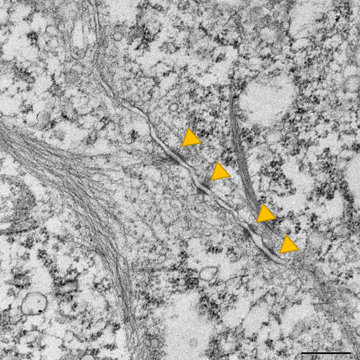 | 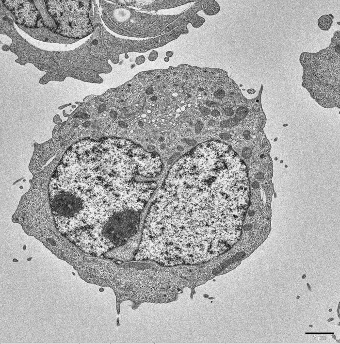 |
| Cell to cell Contact | Cell |
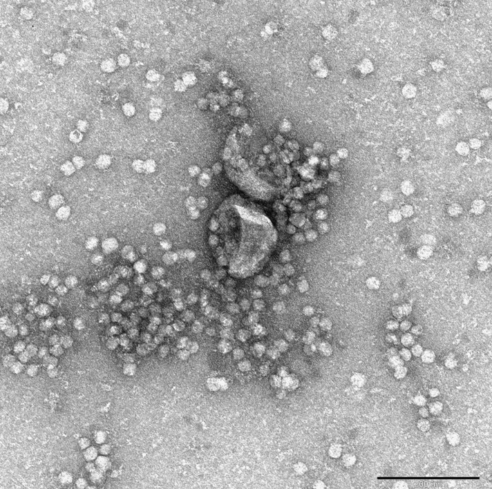 | 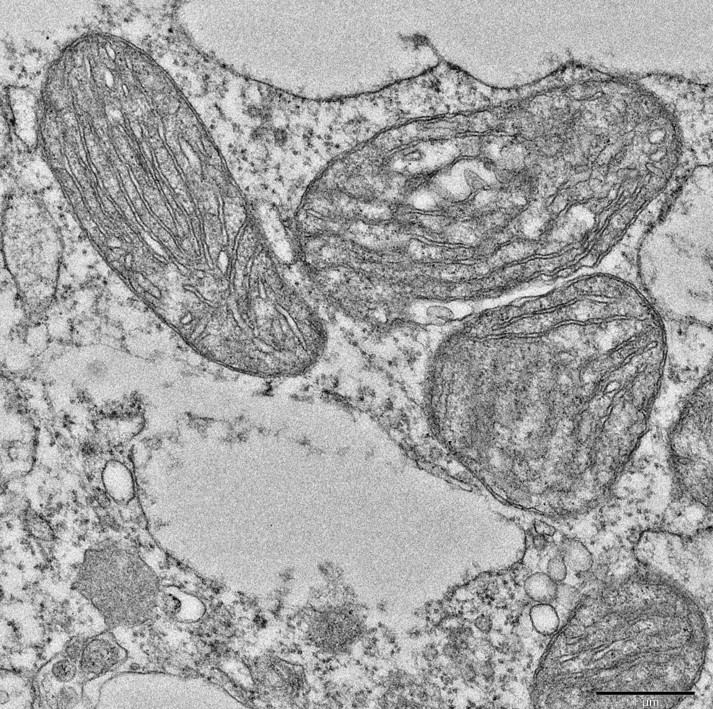 |
| Exosomes | Mitochondria |
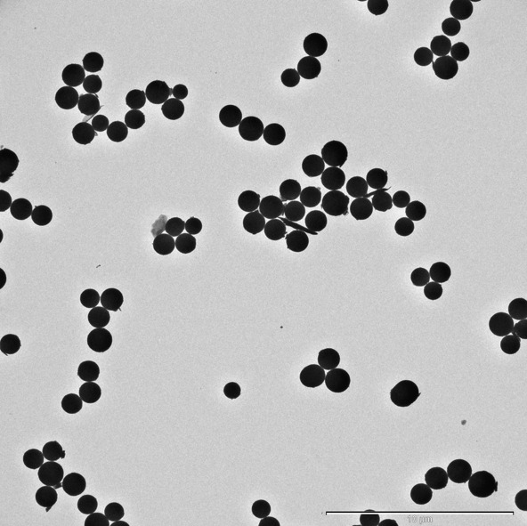 | 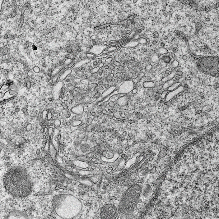 |
| Nanoparticles | Golgi Complex |