

Digital Imaging

The Digital Imaging Facility is a centralized core facility that provides advanced microscopy and image analysis services to students and researchers across the institute. By integrating state-of-the-art imaging technologies with powerful image analysis tools, the facility enables researchers to visualize, quantify, and gain deeper insights into complex biological structures and processes.
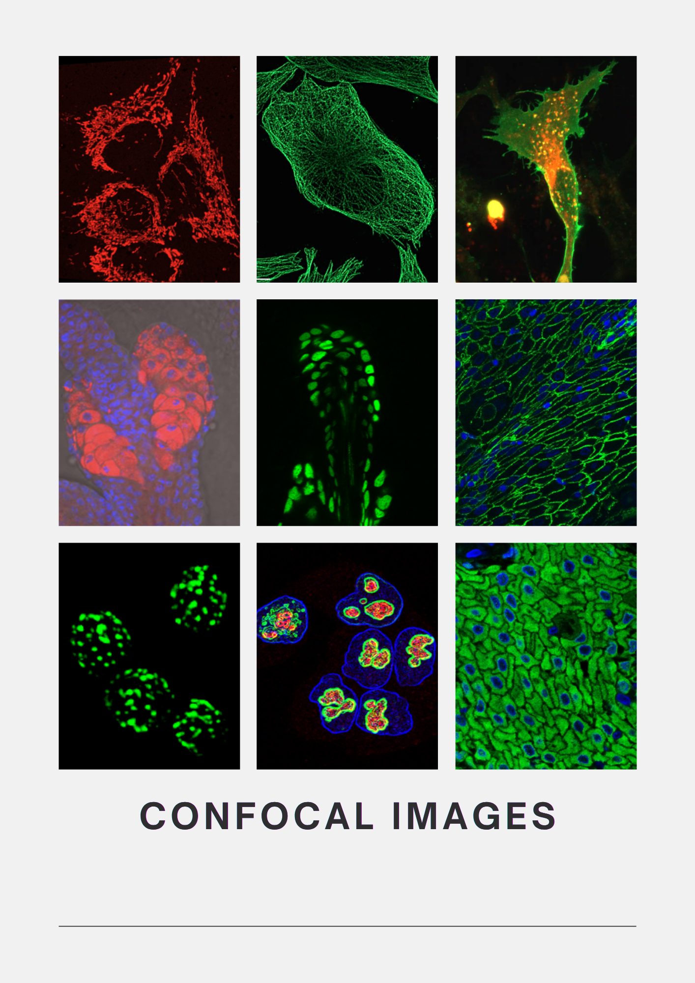
The facility offers a comprehensive range of imaging platforms, including five widefield microscopes, three confocal microscopes all with dedicated live-cell imaging capabilities, and the Incucyte Live-Cell Imaging and Analysis System for long-term monitoring of living cells. Complementing these systems are dedicated high-performance image analysis workstations equipped with software such as Imaris and Arivis, enabling sophisticated 3D reconstruction, visualization, deconvolution, quantitative analysis, and interpretation of multidimensional imaging datasets.
The facility serves more than 18 research laboratories and over 50 active users, with the confocal systems operating at near full capacity. Extended operating hours and weekend access for certified users provides greater flexibility. Beyond our institution, we also welcome researchers from external organizations to utilize the facility for imaging their samples.
Our imaging platforms support a wide range of biological samples, from cultured cells and tissues to whole organisms, enabling diverse research applications. These include protein localization and colocalization, live-cell imaging, drug uptake and response studies, apoptosis, cell migration and invasion, tumour biology, and many other areas of biomedical research. The facility also offers expertise in advanced imaging applications such as FRET (Förster Resonance Energy Transfer) and FRAP (Fluorescence Recovery After Photobleaching), allowing researchers to study protein interactions, molecular mobility, and cellular dynamics with high spatial and temporal resolution.
The Digital Imaging Facility is more than a collection of instruments; it is a center for learning. We provide user training and certification programs, guidance on sample preparation and experimental design, and technical support throughout the imaging process. Our team works closely with our facility users to recommend the most appropriate microscopy techniques and instruments based on their specific research needs, helping them achieve the best possible imaging results.
- Mrs. Vaishali Kailaje, Scientific Assistant,’F’ , Facility In charge
- Mrs. Tanuja Durve, Scientific Assistant, ‘E’
- Ms. Rajashree Sawant, Scientific Assistant, ‘B’
Facility Team:
- Dr. Sorab Dalal, Scientific Officer, ‘G’, Deputy Director, CRI
- Dr. Rohan Khadilkar, Scientific Officer, ‘E’
Facility Board Members:
- Mr. Uday Dandekar. Engineer, ‘H’
Facility Reporting Officer:
- Ms. Shivali Mishra
- Mr. Sourav Chakraborty
Student’s representatives:
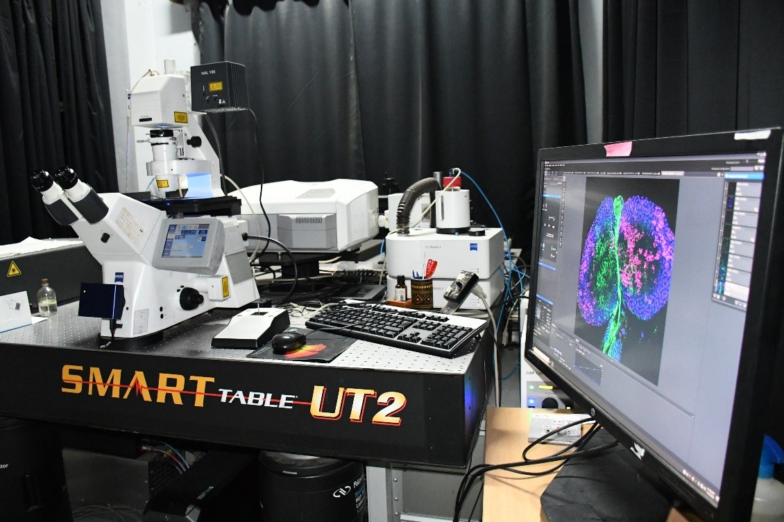
ZEISS LSM 980 AS2 Confocal
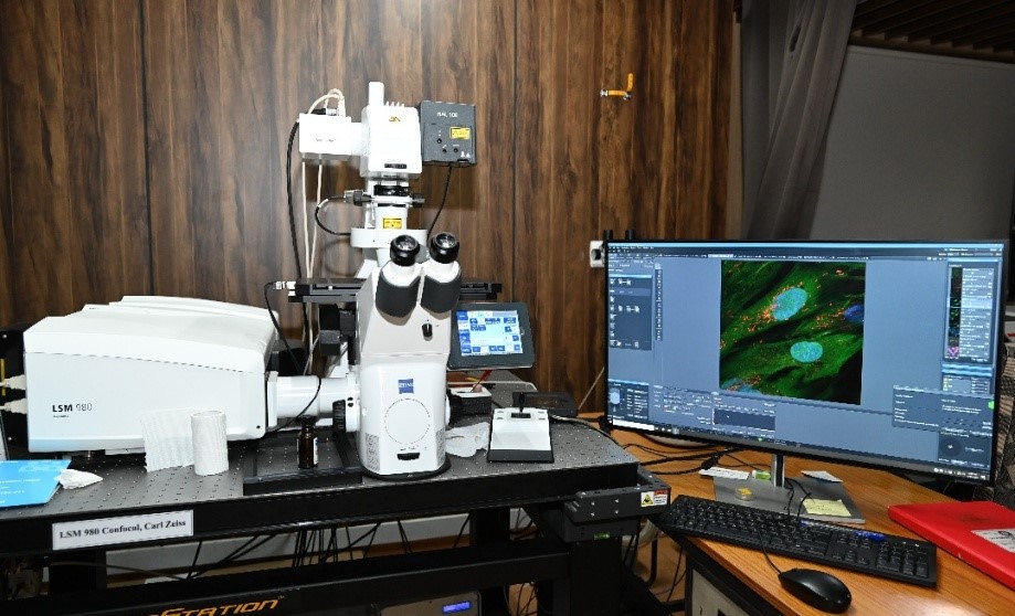
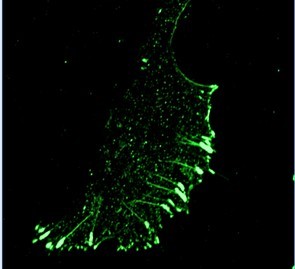
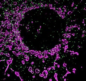
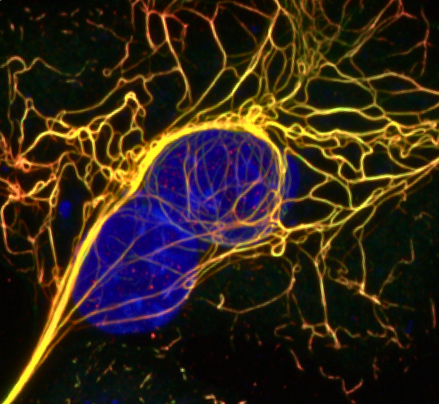
The ZEISS LSM 980 with Airyscan 2 is a next-generation laser scanning confocal microscope designed for high-sensitivity, and super-resolution fluorescence imaging. Featuring the innovative Airyscan 2 detector, the system captures significantly more light and spatial information than conventional confocal microscopes, delivering enhanced resolution, superior signal-to-noise ratio, and improved imaging speed with minimal phototoxicity. Integrated with automated stage control, Definite Focus technology, spectral detection, and live-cell imaging capabilities, the LSM 980 is an ideal platform for advanced biological research.
| Objectives | 10x/0.3NA, 20x/0.7NA DIC, 40x/1.25NA, 63x/1.4NA Oil, 100x/1.4NA Oil |
| Lasers | 405nm, 488nm, 514nm, 561nm, 594nm, 639nm (all solid-state lasers) |
| Detectors | Four-channel GaAsP PMTs, Two-channel Multi-Alkali PMTs (6 spectral channels), Airyscan 2 detector, and Transmitted Light PMT |
| Software | ZEISS ZEN Blue (Version 3.x) |
| Detection Modes | Spectral imaging, Airyscan Super-Resolution, Multiplex imaging, Transmitted light imaging |
CONFOCAL SYSTEMS Nikon AX Confocal
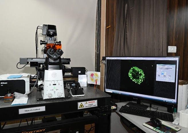
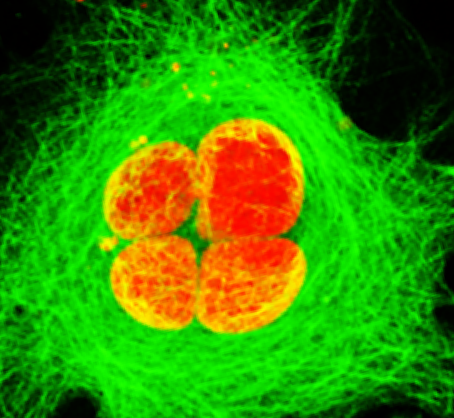
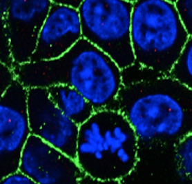
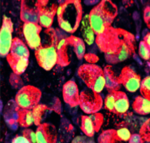
The Nikon AX Confocal Microscope is a state-of-the-art laser scanning confocal imaging system engineered for high-speed, high-resolution, and large field-of-view imaging. Built on the Nikon Ti2-Eclipse fully motorized inverted microscope, with 25 mm ultra-large field-of-view galvo scanner, up to 8192 × 8192-pixel image acquisition, imaging speeds up to 10 fps, 360° scan field rotation, Perfect Focus System (PFS) for long-term live-cell imaging, environmental incubator for 35 mm dishes and multiwell plates.
| Objectives | 10X/0.3NA, 20X/0.7NA DIC, 40X/1.25NA Si (silicon oil),60X/1.4NA Oil, 100X/1.4NA Oil |
| Lasers | 405nm, 488nm, 561nm, 640nm (all solid-state lasers) |
| Detectors | 4 GASP detectors, one Transmitted Light Detector. Both Channel mode (filter based) and Spectral mode imaging possible |
| Software | NIS Elements AR (Ver.5.4.2) is used both as acquisition and analysis software. Acquisition with XY Overview enables selection of region of interest from a large field of view. Workstation PC includes AI based software innovations like, Auto signal.ai and Denoise.ai and deconvolution to improve resolution. |
| Imaging Modes | Brightfield, Phase contrast, DIC, Fluorescence |
LSM780 Confocal
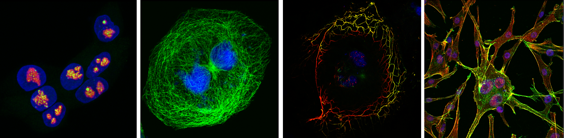
| Features | AxioObserver Z.1 Inverted microscope attached to confocal scan head, Definite Focus - Autofocus System, Motorized XY Stage, Excite Metal Halide Light Source, RGB filter sets for Epi-fluorescence visualization. The scanning field size can go up to 6144 x 6144 pixels. Rotation of the scan field over 360º is possible. A stage mount incubation chamber (maintained at temperature 37ºC and 5% CO2) can be attached for live cell imaging. |
| Objectives | 10x/0.45NA Air (PH1), 20x/0.8NA Air (PH2), 40x/1.3NA Oil, 63x/1.4NA Oil, 100x/1.4NA Oil. |
| Lasers | 405nm Diode, Multiline Argon laser 458nm, 488nmn, 514nm lines, DPSS 561nm, HeNe 594m, HeNe 633nm. |
| Detectors | The system consists of 3 detectors, 2PMTs and a 34 Channel Spectral Array, all of them can be used for fluorescence and reflection imaging. An additional PMT for transmitted light allows for simultaneous detection of fluorescence and bright field/DIC images. |
| Software | The system is controlled under LSM Software, ZEN 2.3 (Black) Ver. 14.1. A Workstation PC for analysis with the same software is available, it enables in quantitating length / area / mean fluorescence intensity, colocalization coefficients, assigning different LUTs etc. and exporting of images. Additionally, it helps in FRAP analysis. |
- High-resolution fluorescence imaging of cells and tissues
- Optical sectioning and three-dimensional (3D) reconstruction of cells, tissues, organoids spheroids etc.
- Live-cell and short and long-term time-lapse imaging to study migration, division, autophagy and dynamics of organelles and proteins.
- Multi-color fluorescence imaging
- Protein localization and intracellular trafficking studies
- Colocalization and interaction analysis of biomolecules (FRET)
- Fluorescence Recovery After Photobleaching (FRAP) to study mobility and diffusion of proteins.
- Cell morphology and cytoskeletal organization studies
- Organelle imaging (mitochondria, nucleus, lysosomes, Golgi, ER, etc.)
- Cancer biology and tumor microenvironment research
- Drug screening and pharmacological studies
- Large-area tissue imaging using tile scanning
- Quantitative fluorescence intensity measurements
- Biomaterial and scaffold imaging
- Reflection imaging of non-fluorescent specimens
WIDEFIELD SYSTEMS - Axio Imager Z.1 Upright Microscope, Carl Zeiss
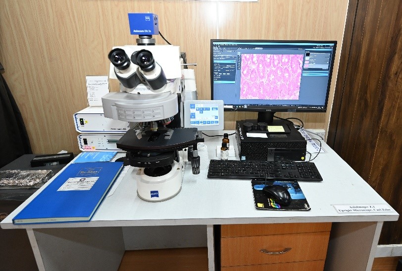
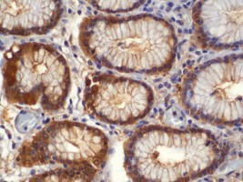
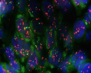
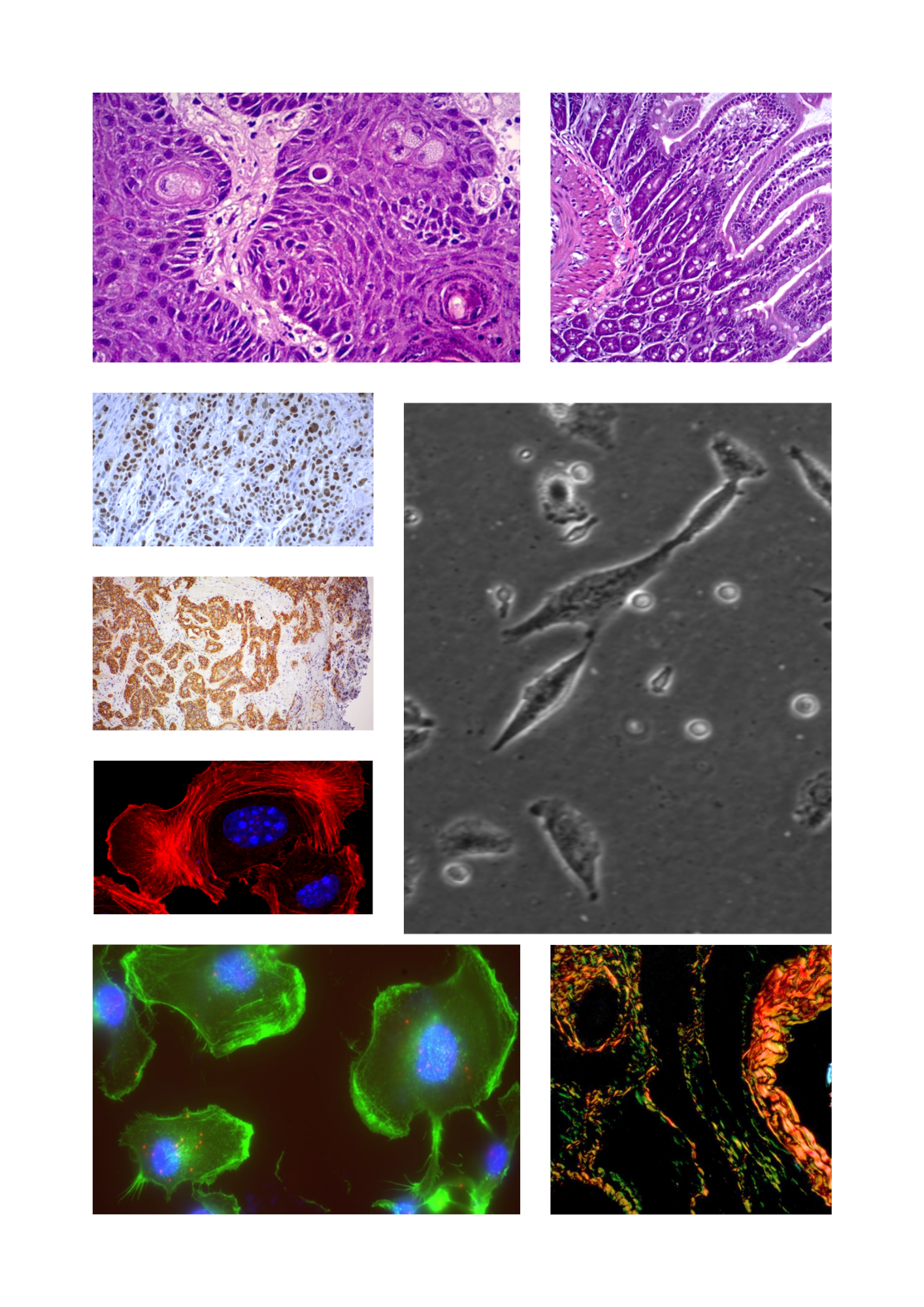
The AxioImager Z.1 is an upright fluorescence microscope used for imaging of cells, tissues, and diverse biological specimens. Partially motorized components, equipped with ZEISS optics - 6 position emission filter wheel and objective turret.
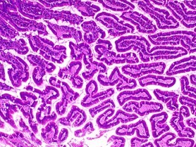
| Objectives | 5X/0.12NA Air, 10X/0.45NA Air (PH1), 20X/0.8NA Air (PH2), 40X/1.3NA Oil, 40X/0.95NA Air, 63X/1.4NA Oil, 100X/1.4NA Oil. |
| Detectors | Equipped with a colored camera Axiocam ERc 5s, Carl Zeiss. |
| Software | Zen3 PRO (Blue Edition) Ver.3.0 |
| Imaging Modes | Brightfield, Phase Contrast and Fluorescence |
| Applications | H&E-stained tissues, Immunohistochemistry (IHC), Fluorescence imaging of cells and tissue sections. |
BX63, Upright Microscope, Olympus
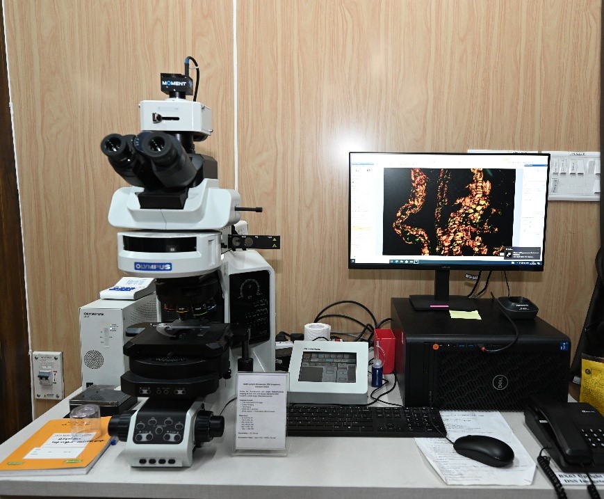
The Olympus BX63 is a partially motorized upright microscope used for imaging of biological specimens - cells, tissues, organism etc. with 8 position emission filter wheel, LED as illumination sources for fluorescence light.
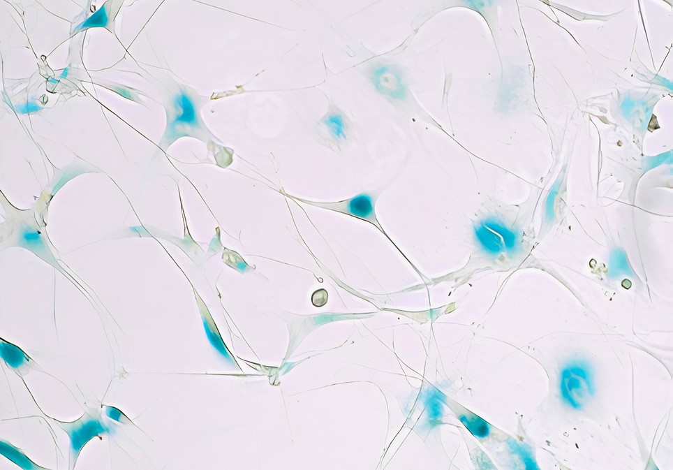
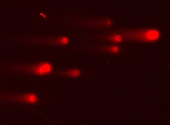
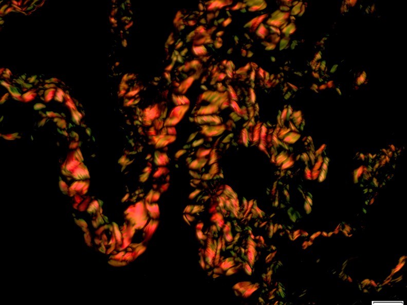
| Objectives | 10X/0.3NA Air (PH1), 20X/0.7NA Air (PH2), 40X/0.9NA Air, 60X/1.4NA Oil, 100X/1.4NA Oil. |
| Detectors | Monochrome (Moments, Teledyne Photometrics) Colored camera (DP23, Olympus) |
| Software | Cell Sens Dimension Software (Ver. 3.2) |
| Imaging Modes | Brightfield, Fluorescence, Polarization microscopy |
| Applications | H&E stained histopathology tissues, Immunohistochemistry, fluorescently labelled cells and tissue, cells and tissues under polarized light |
IX73 Inverted Microscope, Olympus
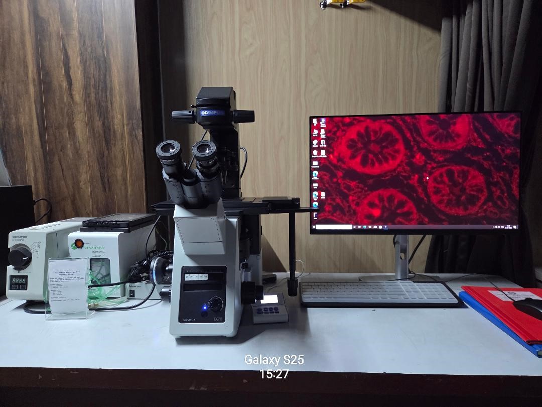
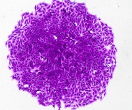
The Olympus IX73 is a compact inverted fluorescence microscope designed for routine cell culture observation and basic fluorescence imaging. Equipped with phase contrast, fluorescence illumination, and a color digital camera
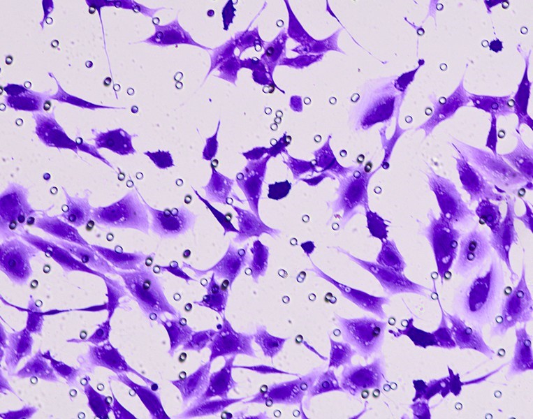
| Objectives | 10X/0.30 Air, 20X/0.70 Air, 40X/0.65 Air |
| Detectors | Colored camera DP28, Olympus |
| Software | Cell Sens Dimension Software (Ver. 3.2) |
| Imaging Modes | Brightfield, Phase contrast, Fluorescence |
| Applications | Cell culture observation, Fluorescence microscopy, Phase contrast imaging, Cell morphology analysis, Cell motility and invasion assays Transfection efficiency assessment |
Axio Observer Inverted Microscope, Carl Zeiss
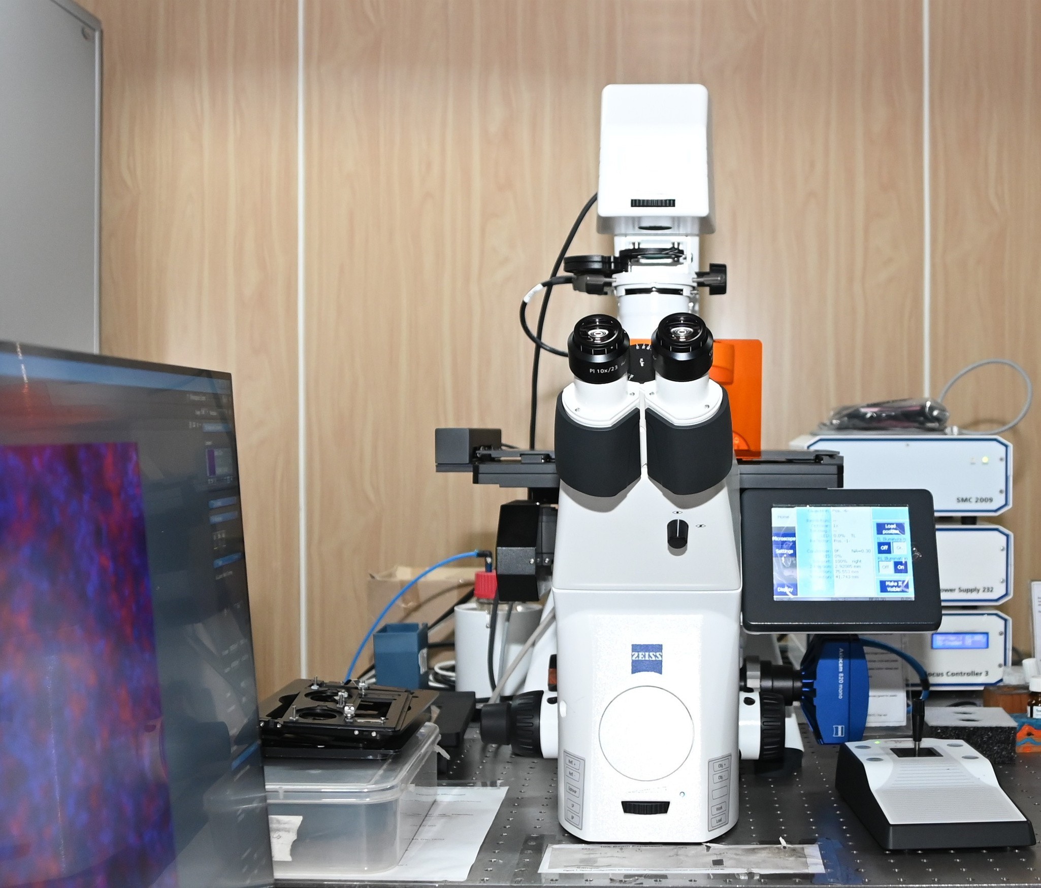
The Carl Zeiss Axio Observer is a fully motorized inverted fluorescence microscope used for advanced live-cell and fluorescence imaging. Featuring an environmental chamber, Colibri LED illumination and Definite focus for z-drift compensation.
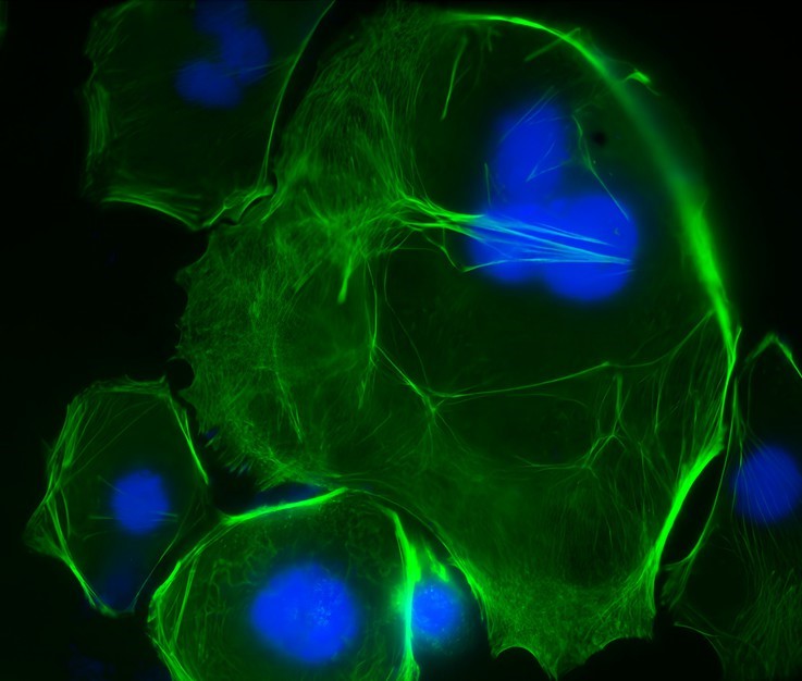
| Objectives | 10X.45 Air (PH1), 20X0.80 Air (PH2), 40X0.95 Air (DIC) |
| Detectors | High-sensitivity AxioCam 802 monochrome camera |
| Software | ZEN Pro 2.0 (Blue Edition) software |
| Imaging Modes | Brightfield, Phase Contrast, DIC, Fluorescence, Tile Scan, Time-lapse and Multidimensional imaging |
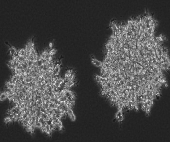
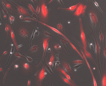
| Applications | Time-lapse imaging, Tile scan imaging, Transfection efficiency assessment, Multidimensional imaging (XYZCT), Cell migration and proliferation studies |
Nikon Eclipse Ti Inverted Microscope
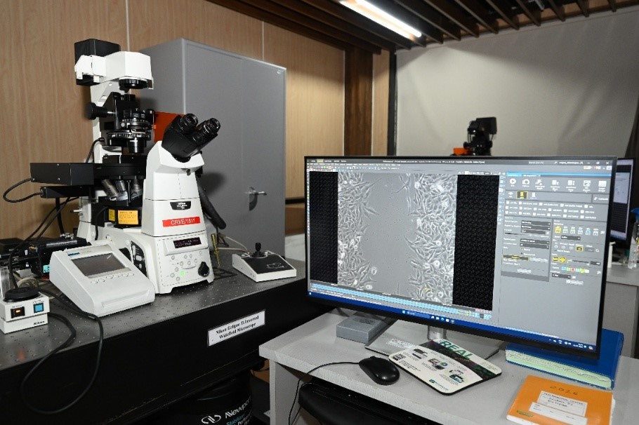
Nikon Eclipse Ti Inverted Microscope is a Live-cell and fluorescence imaging system. Equipped with an environmental chamber that maintains physiological conditions (37°C and 5% CO₂), the system enables long-term imaging of living cells. It has the Perfect Focus for Z drift compensation.
| Objectives | 10X0.30 Air (PH1), 20X0.70 DIC, 20X0.70 Air (PH2) |
| Detectors | 2 Monochrome camera - ORCA-Flash4.0 and Andor Clara |
| Software | NIS Elements AR software (Ver.4.2) |
| Imaging Modes | Brightfield, Phase Contrast, DIC, Fluorescence, Tile Scan, Time-lapse |
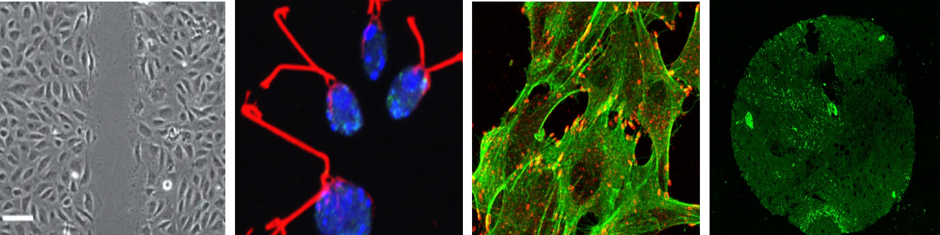
| Applications | Time-lapse imaging, Wound healing assays, Fluorescence microscopy Cell viability and proliferation , Transfection efficiency studies, Phase Contrast and DIC imaging, Cell motility and invasion assays. |
Incucyte
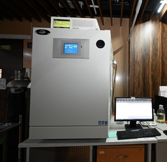
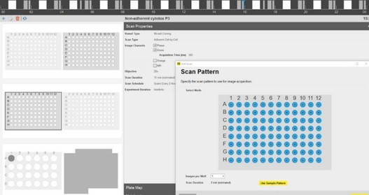
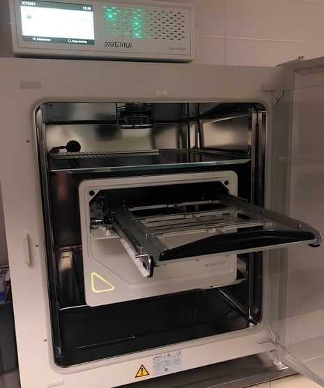
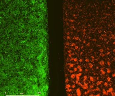
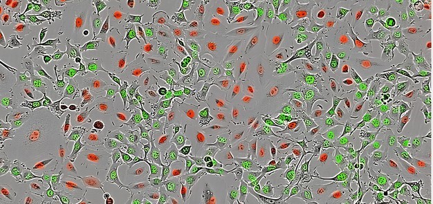
This is a widefield inverted live cell system engineered with long term high throughput imaging in mind. Housed inside a tissue culture incubator, the imaging module maintains the most stable imaging conditions possible (37ºC temperature, humidity and 5% CO2). The system comes with a selection of inserts allowing you to image your cells in a range of vessels (ranging from 35mm dishes to 384 well plates). It automatically captures and analyses images of living cells in real time for days, weeks, or months. It has a optical design wherein the cells remain stationary while optics move. Multiple imaging modes – HD phase plus red and green fluorescence. It can accommodate multiple users at a time with different experiment set ups and configurations
| Objectives | 4X, 10X, 20X |
| Detectors | CMOS camera |
| Imaging Modes | Phase contrast, fluorescence (Red and Green only) |
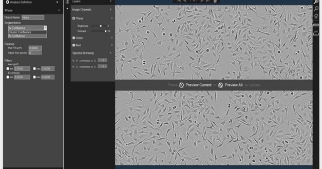
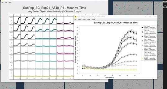
| Applications | The acquisition software comes with automated analysis tools to aid with high throughput analysis, such as object counting, scratch wound closure, and confluence measurements. Results are also displayed as plate maps making it easy to see instantly how parameters change between different wells/treatments. You can visualize and validate results by exporting them in the form of images / movies/ data tables. Analysis modules (Essen Incucyte 2020A) available with the system are, Basic Analyzer, Scratch Wound, Chemotaxis, etc. |
Dedicated analysis workstations equipped with Imaris, Arivis Pro
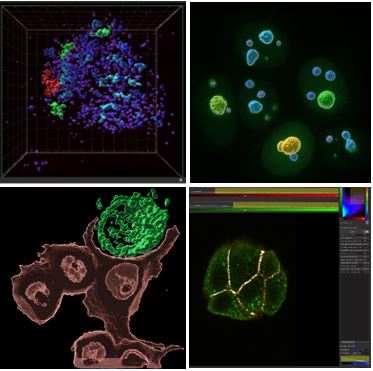
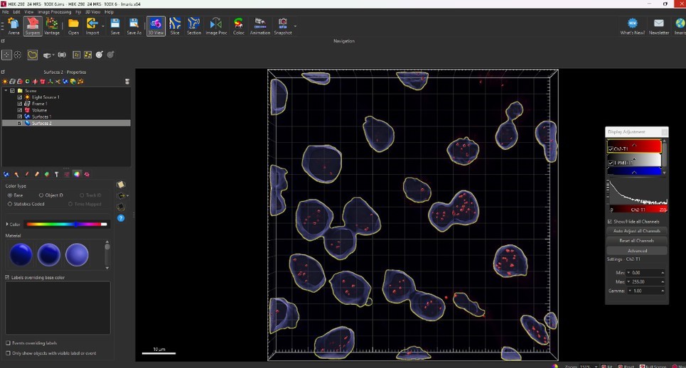
Imaris for advanced 3D visualization, segmentation, cell tracking, and quantitative image analysis.
Arivis Pro for AI-assisted segmentation, analysis of large image datasets, multidimensional visualization, and automated workflows.
 |  |
Advanced Microscopy
Confocal laser scanning microscopy and improved-resolution imaging of subcellular structures with Airy Scan hardware. Multi-channel fluorescence imaging, Spectral imaging and spectral unmixing
Widefield fluorescence microscopy - Brightfield, phase-contrast, and DIC imaging, Tile scan - large-area imaging.
Time-lapse imaging - long-term live-cell imaging under controlled temperature and CO₂ conditions with automated multilocation and multichannel imaging.
Image Processing
Image deconvolution, noise reduction and AI-assisted denoising, spectral unmixing,3D reconstruction and rendering, image stitching, movie generation and image annotation
Quantitative Image Analysis
2D, 3D, and 4D image visualization, cell segmentation and object detection, colocalization analysis, fluorescence intensity measurements, surface, volume, and morphology quantification, AI-assisted image segmentation and analysis.
Functional Imaging
- Fluorescence Recovery After Photobleaching (FRAP) / FRET (Förster Resonance Energy Transfer)
- Cell migration and wound-healing assays
- Chemotaxis and proliferation studies
- Confluence and viability analysis using Incucyte
User Training and Technical Support
- Instrument operation training
- Experimental planning and consultation
- Assistance in optimizing imaging protocols for diverse biological samples
- Selection of appropriate imaging techniques
- Assistance with image acquisition and optimization
- Image analysis guidance
- Troubleshooting and technical support
Data Management
We are happy to assist you with your microscopy and image analysis requirements. Feel free to contact us by email or phone for any queries related to the facility or our services.
In-house Certified Users
Certified in-house users can access the facility independently after regular working hours, as well as on weekends and public holidays, subject to facility guidelines and instrument availability.
External Users
Researchers from external institutes are welcome to use the facility. Please email us with a brief description of your project, your imaging requirements, and the microscope(s) you wish to use. Our team will guide you in selecting the most suitable system and assist with scheduling your experiments.
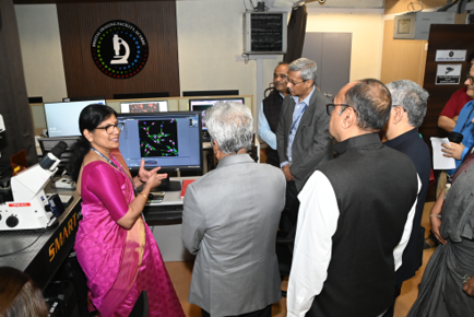
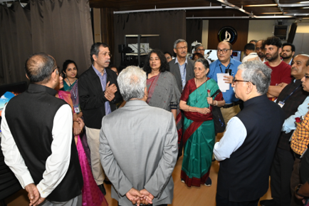
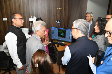
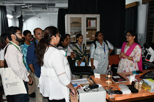
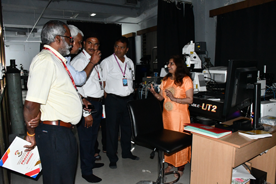
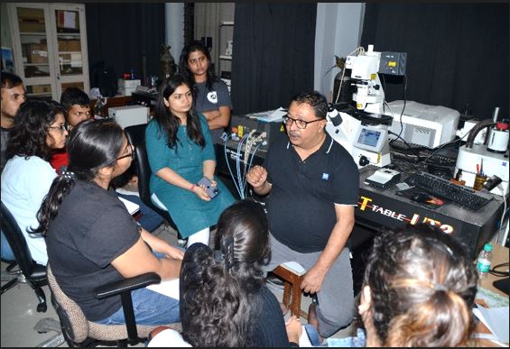
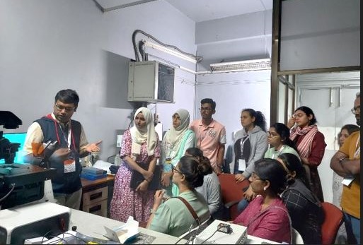
INAUGURATION OF DIGITAL IMAGING FACILITY (REIMAGINED AND RENOVATED)


SCIENCE DAY
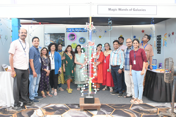
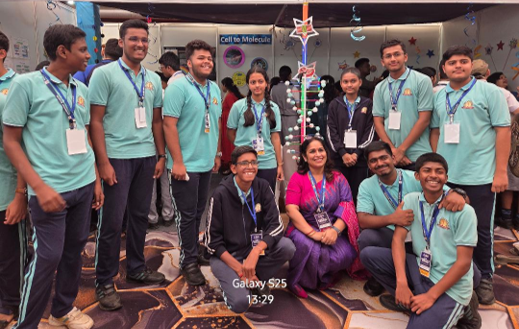
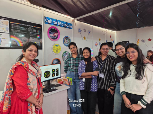
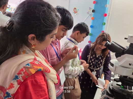
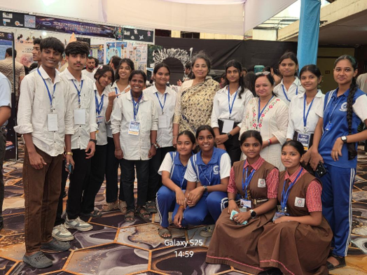
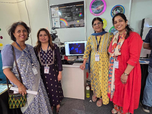
Workshop & Visits
2026
- Mehta D, Paradkar A, Rekhi B, Nayak P, Mohanty B, Chaudhari P, Gera P, Rane S, Chaturvedi P, Waghmare SK. Histone methylation defines c-Jun/Sox2/Hif1α axis that controls stemness and tumor progression in squamous cell carcinoma. Nature Communications. PMID: 42045185
- Dey P, Bishnu A, Patel J, Mishra S, Gadewal N, Ray P, Gupta S, De A. Hotspot mutations in HER2 interfaces destabilize structure, causing breast cancer treatment failure. Oncogene. 45(3):414-430. PMID: 41392272
- Mehrotra M, Shenoy P, Sakpal A, Chakraborty S, Ray P. p53-R248W evades doxorubicin mediated cell death through collective alteration in TOP2A and CYP1A1 genes in cancer cells. Biochemical and Biophysical Research Communications. 819: 153759. PMID: 42019355
- Manasi Nagare, Monalisa Sahoo, Manju Sengar, Sachin Punatar, Navin Khattry, Anant Gokarn, Bhausaheb Bagal, Hasmukh Jain, Sumeet Mirgh, Sridhar Epari, Tanuja Shet, Trupti Pradhan, Shweta Shirsat, Madan Barkume, Caroline Mathen, Poonam Gera, Rohit Kumar Verma, Elveera Saldanha, Pratik Chandrani, Jitendra Gawde, Shubhada Chiplunkar & Jyoti Kode. Defining alteration in bone marrow mesenchymal stem cells (MSC) from acute myeloid leukemia and exploring cultured MSC-conditioned media as a novel anti-leukemia therapy agent. Cancer Immunology Immunotherapy: 2026 Feb 23;75(3):83
2025
- Anthony C, Harish M, Christie J, Venkatraman P (2025). Novel interaction with PSMD9 regulates DNAJA1 turnover and mitochondrial polarity, Biochemical and Biophysical Research Communications.
- Lonare A, Raychaudhuri K, Shah S, Madhu G, Sachdeva A, Basu S, Thorat R, Gupta S, Dalal SN (2025).14-3-3σ restricts YY1 to the cytoplasm, promoting therapy resistance, and tumor progression in colorectal cancer. International Journal of Cancer.
- Jog E, Jainarayanan AK, La Ferlita A, Chakraborty A, Dalwai A, Yahya S, Shivashankar A, Choudhary BS, Chandramouli A, Kazi M, Jain D, Khapare N, B A, Khan BK, Gera P, Patil P, Thorat R, Verma N, Sehgal L, Saklani A, Kamat SS, Dalal SN, Chaudhary N. Inhibiting de novo lipogenesis identifies a therapeutic vulnerability in therapy-resistant colorectal cancer. Redox Biol. 2025 Feb;79:103458. Epub 2024 Dec 11. PMID: 39705849; PMCID: PMC11729006.
- Choudhary BS, Chaudhary N, Khan BK, Vijan A, Mandal D, Pilankar L, Gawand S, Uttankar P, Sharma M, Shivashankar A, Doloi R, Joshi N, Das M, Dalal SN. LCN2 promotes focal adhesion formation and invasion by stimulating c-Src activation. Journal of Cell Science. 138(11): jcs263663. PMID: 40356520
- Jaiswal MA, Karn A, Das A, Kumari A, Tiwari S, Dalal SN. 14-3-3ε inhibits premature centriole disengagement by inhibiting the activity of Plk1 and separase. Journal of Cell Science. 138(14): jcs263808PMID: 40576425
- Chaudhary N, Mandal D, Choudhary B, Patra S, Jain D, Poonia P, Shaikh S, Tekalkar S, Malvankar S, Shivashankar A, Jog E, Pilankar L, Thorat R, Kailje V, Khanna S, Manna S, Khan B, Jadhav A, Sharma K, Ramu S, Sahoo S, Jolly MK, Dalal SN, Sinha S, Nair N, Ruppin E, Lamballe F, Maina F, Verma N. Low-GPX4 drives a sustained drug-tolerant persister state in TNBC by a targetable adaptive FSP1 upregulation. Redox Biology.
- Joshi P, Bane S, Chaturvedi P, Gera P, Waghmare SK (2025).Establishment and characterization of patient-derived tongue squamous cell carcinoma cell lines. Human Cell. 38(4):102.
- Mehta D, Paradkar A, Nayak P, Rekhi B, Mohanty B, Chaudhari P, Waghmare SK. Establishment and molecular characterization of the novel cutaneous squamous cell carcinoma cell line from advanced-stage Indian patient. Human Cell.38(4):108. PMID: 40413685
- Shenoy PS, Dash S, Joshi D, Rekhi B, Gota V, Ray P. Withaferin A Exerts Cytotoxicity in Single/multidrug-resistant gastric and ovarian cancer cells and tumor xenografts through the AKT-NF-κB-STAT3-survivin axis. Molecular Carcinogenesis
- Chaudhary N, Choudhary BS, Shivashankar A, Manna S, Ved K, Shaikh S, Khanna S, Baar J, Dani J, Sahoo S, Soundharya R, Jolly MK, Verma N. EGFR-to-Src family tyrosine kinase switching in proliferating-DTP TNBC cells creates a hyperphosphorylation-dependent vulnerability to EGFR TKI. Cancer Cell International. 25(1):55. PMID: 39972345.
- Ghosh K, Iyer RK, Sood S, Islam MS, Labad JG, Khadilkar RJ. Genetic perturbation of cellular homeostasis regulates integrated stress response signaling to control Drosophila hematopoiesis. Biology Open. 14(7): bio062046. PMID: 40554752
- Tejashree Mahaddalkar, Archisman Banerjee, Madhura Ketkar, Rahul Thorat, Nilesh Gardi & Shilpee Dutt. Oncogene, volume 44, pages2103–2115 April (2025). Aurora Kinase A and B inhibition abrogates ‘Neosis’, a non-mitotic cell division of GBM residual cells and prevents GBM recurrence.
- Chaudhary N, La Ferlita A, Choudhary BS, Jog E, Kazi M, Yahya S, Dalwai A, Ostwal V, Kumar S, Redkar S, Khapare N, Kailaje V, B A, Gera P, Bal M, Verma N, Thorat R, Saklani A, Sehgal L, Dalal SN (2025). Patient-derived Organoids and Xenografts Uncover Therapeutic Vulnerabilities in Colorectal Signet Ring Cell Carcinomas. Clinical Cancer Research
2024
- Choudhary BS, Chaudhary N, Vijan A, Mandal D, Pilankar L, Gawand S, Khan B, Shivashankar A, Doloi R, Joshi N, Das M, Dalal SN (2024). LCN2 promotes focal adhesion formation and invasion by stimulating Src activation. bioRxiv.
- Tilwani S, Gandhi K, Dalal SN (2024). 14-3-3ε conditional knockout mice exhibit defects in the development of the epidermis. FEBS Letters.
- Roy S, Mehta D, Paradkar A, Chovatiya G, Waghmare SK (2024). Dab2 (Disabled-2), an adaptor protein, regulates self-renewal of hair follicle stem cells. Communications Biology. 7(1): 525.
- Roy S, Mehta D, Sawant S, Waghmare SK (2024). Disabled 2 (Dab2) Regulates Tumour Progression in Skin Squamous Cell Carcinoma
- Phadte P, Bishnu A, Dey P, M M, Mehrotra M, Singh P, Chakrabarty S, Majumdar R, Rekhi B, Patra M, De A, Ray P (2024). Autophagy-mediated ID1 turnover dictates chemo-resistant fate in ovarian cancer stem cells. Journal of Experimental and Clinical Cancer Research. 43(1):222
2023
- Christie J, Anthony CM, Harish M, Mudartha D, Farooqee SBUD, Venkatraman P. The interaction network of the proteasome assembly chaperone PSMD9 regulates proteostasis. FEBS Journal.
- Choudhary B, Chaudhary N, Shah M, Dwivedi N, Smitha PK, Das M, Dalal SN. Lipocalin 2 inhibits actin glutathionylation to promote invasion and migration. FEBS Letters.
- Pratiksha Mahadik, Sejal Patwardhan. ECM stiffness-regulated exosomal thrombospondin-1 promotes tunneling nanotubes-based cellular networking in breast cancer cellsArchives of Biochemistry and Biophysics, 742, 109624, (2023) PMID: 37146866
- Joshi P, Waghmare S. Molecular signaling in cancer stem cells of tongue squamous cell carcinoma: Therapeutic implications and challenges. World Journal of Stem Cells 15(5): 438-452.
- Mehta D, Shaikh S, Mohanty B, Chaudhari P, Waghmare SK (2023). Secretory phospholipase (sPLA2-IIA) regulates breast cancer stem cells differentiation and metastatic potential. Biochemical and Biophysical Research Communication. 677:98-104. PMID: 37566923.
- Navarange SS, Bane SM, Mehta D, Shah S, Gupta S, Waghmare SK (2023). Epithelial-to-mesenchymal transition status correlated with ultrastructural features, and TP53 mutation in patient-derived oral cancer cell lines. Molecular Biology Reports. 50(10):8469-8481. PMID: 37639153
- Nazia Chaudhary, Bhagya Shree Choudhary, Anusha Shivashankar, Subhakankha Manna, Khyati Ved, Shagufa Shaikh, Sonal Khanna, Jeetnet Barr, Jagruti Dani, Nandini Verma. EGFR-to-Src family tyrosine kinase switching in proliferating-DTP TNBC cells creates a hyperphosphorylation-dependent vulnerability to EGFR TKI. bioRxiv 2023.07.17.549374;
- Kode, Jyoti; Maharana, Jitendra; Dar, Asif; Mukherjee, Shayanti; Gadewal, Nikhil; Sigalapalli , Dilep; Kumar, Satyanshu; Panda, Debashis; Ghosh , Soumyajit ; Keshry , Supriya ; Mamidi , Prabhudutta ; Chattopadhyay , Soma ; Pradhan, Trupti; Kailaje, Vaishali; Inamdar, Sunil; Gujjarwar , Vidula. 6-Shogaol exhibits anti-viral and anti-inflammatory activity in COVID-19 associated inflammation by regulating NLRP3 inflammasome. ACS Omega 8, 2, 2618–2628
- Low levels of CD26 on certain cellular subtypes of donor harvest is associated with better clinical outcomes post allogeneic stem cell transplantation through regulation of NF-κB pathway and pro-inflammatory cytokines. International Immunopharmacology. 125(Part A): 111054.PMID: 37890379. Kandekar S, Punatar S, Khattry N Gokarn A, Jindal N, Mirgh S, Chichra A, Tembhare P, Rane P, Gawade J, Mathew L, Patil A, Chiplunkar S, Kode J (2023).
- RajeshKumar Meher , Jitendra Maharana , Jeshma Kovvuri , K.Nirmal Kumar , Trupti Pradhan , Vaishali Kailaje , Arvind Ingle , Subrata Sen , Madan Barkume, Meena Patkar, Anand Vaibhaw, ShowkatAhmad Mir, Arpita Ghosh, Sanjeev Kamte, Ahmed Kamal j, Jyoti Kode. A Phenstatin-based indole-chalcone hybrid as novel anti-inflammatory agent targeting the NLRP3 Inflammasome: Leads from computational and preclinical studies
- Dey P, Gadewal N, De A. Pathogenic HER3 dimerization domain mutations create a structural bias towards un-conventional EGFR-HER3 signalling axis in breast cancer. International Journal of Biological Macromolecules.
- Mehta D, Shaikh S, Mohanty B, Chaudhari P, Waghmare SK (2023). Secretory phospholipase (sPLA2-IIA) regulates breast cancer stem cells differentiation and metastatic potential. Biochemical and Biophysical Research Communication. 677:98-104. PMID: 37566923.
2022
- Mal A, Bukhari AB, Singh RK, Kapoor A, Barai A, Deshpande I, Wadasadawala T, Ray P, Sen S, De A. EpCAM-Mediated Cellular Plasticity Promotes Radiation Resistance and Metastasis in Breast Cancer. Frontiers in Cell Developmental Biology. 8:597673 2021 PMID: 33490064
- Bose, A., Modi, K., Dey, S., Dalvi, S., Nadkarni, P. Sudarshan, M., Tapas K. Kundu, T.K., Venkatraman, P., and Dalal, S.N. 14-3-3γ prevents centrosome duplication by inhibiting NPM1 function. Genes to Cells, 26, 426-446 2021).
- Sarika Tilwani, Karan Gandhi, Satya Narayan, Sri Rama Koti Ainavarapu, Sorab Nariman Dalal (2021).Disruption of desmosome function leads to increased centrosome clustering in 14-3-3γ-knockout cells with supernumerary centrosomes.
- Raghavan, R., Koyand, N., Beher, R., Chetlangia, N., Ramadwar, M., Pawade, S., Thorat, R., van Hengel, J., Sklyarova, T., van Roy, F., and Dalal, S.N. (2022). Plakophilin3 loss leads to increased adenoma formation and rectal prolapse in APCmin mice. Biochem. Biophys. Res. Commun.586, 14-19
- N. Chaudhary, N. Joshi, R. Doloi, A. Shivashankar, R. Thorat and S. N. Dalal. (2022). Plakophilin3 loss leads to an increase in autophagy and radio-resistance. Biochem Biophys Res Commun.620, 1-7.
- Singh, Bhawna, et al. The BRCA2 and CDKN1A interacting protein (BCCIP) stabilizes stalled replication forks and prevents degradation of nascent DNA." FEBS letters 596.16 (2022): 2041-2055
- Mulla SW, Venkatraman P. Novel Nexus with NFκB, β-catenin, and RB1 empowers PSMD10/Gankyrin to counteract TNF-α induced apoptosis establishing its oncogenic role. International Journal of Biochemistry & Cell Biology. 146: 106209. PMID: 35378311.
- Mukherjee S, Sakpal A, Mehrotra M, Phadte P, Rekhi B, Ray P (2022). Homo and heterotypic cellular cross-talk in epithelial ovarian cancer impart pro-tumorigenic properties through differential activation of the Notch3 pathway. Cancers. 14(14): 3365.
- Mishra SK, Dhadve AC, Mal A, Reddy BPK, Hole A, CM Krishna, Ray P, Srivastava R, De A. Photothermal therapy (PTT) is an effective treatment measure against solid tumors which fails to respond conventional chemo/radiation therapies in clinic. Biomaterials Advances. 143:213153. PMID: 36343390
- Mukherjee S, Sharma S, Soni V, Joshi A, Gaikwad A, Bellare J, Kode J. Improved osteoblast function on titanium implant surfaces coated with nanocomposite Apatite-Wollastonite-Chitosan- an experimental in-vitro study. Journal of Materials Science: Materials in Medicine. 33(3):25. PMID: 35190908.
2021
- Sheikh Burhan Ud Din Farooqee, Joel Christie, Prasanna Venkatraman. PSMD9 ribosomal protein network maintains nucleolar architecture and WT p53 levels, Biochemical and Biophysical Research Communications, 2021
- Chaudhary N, Choudhary BS, Shah SG, Khapare N, Dwivedi N, Gaikwad A, Joshi N, Raichanna J, Basu S, Gurjar M, Smitha PK, Saklani A, Gera P, Ramadwar M, Patil P, Thorat R, Gota V, Dhar SK, Gupta S, Das M, Dalal SN. Lipocalin 2 expression promotes tumor progression and therapy resistance by inhibiting ferroptosis in colorectal cancer, Int J Cancer.
- Bose A, Modi K, Dey S, Dalvi S, Nadkarni P, Sudarshan M, Kundu TK, Venkatraman P, Dalal SN. 14-3-3γ prevents centrosome duplication by inhibiting NPM1 function. Genes Cells, 26(6):426-446. PMID: 33813791
- Mal A, Bukhari AB, Singh RK, Kapoor A, Barai A, Deshpande I, Wadasadawala T, Ray P, Sen S, De A. EpCAM-Mediated Cellular Plasticity Promotes Radiation Resistance and Metastasis in Breast Cancer. Frontiers in Cell Developmental Biology. 8:597673 2021 PMID: 33490064
2020
- Ramchandra V Amnekar, Shafqat A Khan, Mudasir Rashid, Bharat Khade, Rahul Thorat, Poonam Gera, Shailesh V Shrikhande, Duane T Smoot, Hassan Ashktorab, Sanjay Gupta. Histone deacetylase inhibitor pre-treatment enhances the efficacy of DNA-interacting chemotherapeutic drugs in gastric cancer (2020). World J Gastroenterol 2020 February 14; 26(6): 598-613
- Sanket G. Shah, Tripti Verma, Mudasir Rashid, Nikhil Gadewal, Sanjay Gupta. Histone H2A isoforms: Potential implications in epigenome plasticity and diseases in eukaryotes (2020). J of Biosciences 45:4
- Asmita Sharda, Mudasir Rashid, Sanket Shah, Ajit Kumar Sharma, Saurav Raj Singh, Poonam Gera, Murali Krishna Chilakapati, Sanjay Gupta. Elevated HDAC activity and altered histone phospho-acetylation confer acquired radio-resistant phenotype to breast cancer cells (2020) Clinical Epigenetics. 12:4
- Sanket G. Shah, Tushar Mandloi, Pooja Kunte, Abhiram Natu, Mudasir Rashid, Divya Reddy, Nikhil Gadewal & Sanjay Gupta. HISTome2: a database of histone proteins, modifiers for multiple organisms and epidrugs. Epigenetics & Chromatin; 13: 31 (2020)
- Mukherjee S, Agarwal M, Bakshi A, Sawant S, Thomas L, Fujii N, Nair P, Kode J. Chemokine SDF1 Mediated Bone Regeneration Using Biodegradable Poly(D, L-lactide-co-glycolide) 3D Scaffolds and Bone Marrow-Derived Mesenchymal Stem Cells: Implication for the Development of an “Off-the-Shelf” Pharmacologically Active Construct. Biomacromolecules
- Kode J, Kovvuri J, Nagaraju B, Jadhav S, Barkume M, Sen S, Kasinathan NK, Chaudhari P, Mohanty BS, Gour J, Sigalapalli DK, Kumar CG, Pradhan T, Banerjee M, Kamal A. Synthesis, biological evaluation, and molecular docking analysis of phenstatin based indole linked chalcones as anticancer agents and tubulin polymerization inhibitors. Bioorganic Chemistry.
- Dimri S, Malhotra R, Shet T, Mokal S, Gupta S, De A (2020). Noncanonical pS727 post translational modification dictates major STAT3 activation and downstream functions in breast cancer. Experimental Cell Research. 396(2): 112313. PMID: 33002501
- Rathod M, Kelkar M, Valvi S, Salve G, De A (2020). FOXA1 regulation turns benzamide HDACi treatment effect specific in breast cancer promoting NIS gene mediated targeted radioiodine therapy. Molecular Therapy: Oncolytics. 19: 93-104. PMID: 33102692
- Mal A, Dey P, Hayes RM, McCarthy JV, Ray A, De A (2020). In Silico Identification of Potential Phosphorylation in the Cytoplasmic Domain of Epithelial Cell Adhesion Molecule. ACS Omega. 5(48): 30808-30816. PMID: 33324790
- Ghorai, A., Mahaddalkar, T., Thorat, R. and Dutt, S. (2020). Sustained inhibition of PARP-1 activity delays glioblastoma recurrence by enhancing radiation-induced senescence. Cancer Lett. 490, 44-53.
2019
- Harish M, Kannan S, Puttagunta S, Pradhan MR, Verma CS, Venkatraman P. A Novel Determinant of PSMD9 PDZ Binding Guides the Evolution of the First Generation of Super Binding Peptides. Biochemistry. 58(32): 3422-3433, 2019. PMID: 31287951.
- Sahu I, Nanaware P, Mane M, Mulla SW, Roy S, Venkatraman P. Role of a 19S Proteasome Subunit- PSMD10(Gankyrin) in Neurogenesis of Human Neural Progenitor Cells. Int J Stem Cells. 12(3):463-473, 2019. PMID: 31474027
2018
- Mansa Gurjar, Kumar Krishna Raychaudhuri, Snehal Mahadika, Divya Reddy, Apurva Atak, Trupti Shetty Kruthi Rao, Mansi S. Karkhanis, Prajakta Gosavi, Lalit Sehgal, Sanjay Gupta, Sorab N. Dalal. Plakophilin3 increases desmosome assembly, size and stability by increasing expression of desmocollin2, Biochemical and Biophysical Research Communications, Volume 495, Issue 1, 1 January 2018, Pages 768-774
- Vishal SS, Tilwani S, Dalal SN. Plakoglobin localization to the cell border restores desmosome function in cells lacking 14-3-3γ. Biochem Biophys Res Commun. 495(2): 1998-2003. PMID: 29253567
- Basu S, Chaudhary N, Shah S, Braggs C, Sawant A, Vaz S, Thorat R, Gupta S, Dalal SN. Plakophilin3 loss leads to an increase in lipocalin2 expression, which is required for tumour formation. Exp Cell Res. 369(2): 251-265. PMID: 29803740
2017
- Kelkar MG, Thakur B, Derle A, Chatterjee S, Ray P, De A. Tumor suppressor protein p53 exerts negative transcriptional regulation on human sodium iodide symporter gene expression in breast cancer. Breast Cancer Res Treat. 164(3), 603-615.
- Chovatiya, G.L., Sarate, R.M., Sunkara, R.R., Gawas, N.P., Kala, V., and Waghmare, S.K. Secretory phospholipase A2-IIA overexpressing mice exhibit cyclic alopecia mediated through aberrant hair shaft differentiation and impaired wound healing response. Sci Rep 7, 11830-9, PMID: 28912581
- Rathod M, Chatterjee S, Dutta S, Kalraiya R, Bhattacharya D, De A. Mannose glycosylation is an integral step for human NIS localization and function in breast cancer cells (2019). Journal of Cell Science. PMID: 31455607.
2016
- Mukhopadhyay, A., Sehgal, L., Bose, A., Gulvady, A., Senapati, P., Thorat, R., Basu, S., Bhatt, K., Hosing, A. S., Balyan, R., Borde, L., Kundu, T. K., and Dalal, S. N. 2016. 14-3-3γ prevents centrosome amplification and neoplastic progression. Scientific Reports 6, Article number: 26580 (2016)
- Raychaudhuri, K., Chaudhary, N., Gurjar, M., D'Souza, R., Limzerwala, J., Maddika, S., and Dalal, S. N. (2016). 14-3-3sigma loss leads to activation of the epithelial to mesenchymal transition due to the stabilization of c-Jun. J Biol Chem 291: 16068-16081. (2016).
- Rahul M. Sarate, Gopal L. Chovatiya, Vagisha Ravi, Bharat Khade, Sanjay Gupta, Sanjeev K. Waghmare. sPLA2 -IIA Overexpression in Mice Epidermis Depletes Hair Follicle Stem Cells and Induce Differentiation Mediated through Enhanced JNK/c-Jun Activation. Stem Cells, 34(9):2407-17, 2016
2015
- Chatterjee S, Thaker N, De A. Combined 2-deoxy glucose and metformin improves therapeutic efficacy of sodium-iodide symporter-mediated targeted radioiodine therapy in breast cancer cells. Breast Cancer (Dove Med Press). 7:251-65.
- Basu S, Thorat R, Dalal SN. MMP7 is required to mediate cell invasion and tumor formation upon plakophilin3 loss.
- Shafqat A. Khan, Divya R Velga, Sanjay Gupta. Global Histone Posttranslational Modifications and Cancer: Biomarkers for Diagnosis, Treatment and Prognosis? World Journal of Biological Chemistry, 6(4): 333–345, 2015
- Ajit K. Sharma, Shafqat A. Khan, Asmita Sharda, Divya R Velga, Sanjay Gupta. MKP1 phosphatase mediates G1-specific dephosphorylation of H3Serine10P in response to DNA damage Mutation Research, 778:71-9, 2015
- Ajit K. Sharma, Saikat Bhattacharya, Shafqat A. Khan, Bharat Khade, Sanjay Gupta. Dynamic alteration in H3 serine 10 phosphorylation is G1-phasespecific during ionization radiation induced DNA damage response inhuman cells. Mutation Research, 773: 83–91
- Snehal M.Gaikwad ,BhushanThakur, Asmita Sakpal, Ram K.Singh Pritha Ray. Differential activation of NF-κB signaling is associated with platinum and taxane resistance in MyD88 deficient epithelial ovarian cancer cells
2014
- Sehgal L, Usmani, A. Dalal SN, Majumdar, SS. Generation of transgenic mice by exploiting spermatogonial stem cells in vivo. Methods Mol Biol 1194: 327-337.
- Sehgal L, Mukhopadhyay A, Rajan A, Khapare N, Sawant M, Vishal SS, Bhatt K, Ambatipudi S, Antao N, Alam H, Gurjar M, Basu S, Mathur R, Borde L, Hosing AS, Vaidya MM, Thorat R, Samaniego F, Kolthur-Seetharam U, Dalal SN. 14-3-3gamma mediated transport of plakoglobin to the cell border is required for the initiation of desmosome assembly in vitro and in vivo. J Cell Sci 127: 2174-2188
- Shafqat A Khan, Monica Tyagi, Ajit K Sharma, Savio G Barreto, Bhawna Sirohi, Mukta Ramadwar, Shailesh V Shrikhande, Sanjay Gupta. Cell-type specificity of β-actin expression and its clinicopathological correlation in gastric adenocarcinoma. World Journal of Gastroenterology, 20(34):12202-11. 2014
- Ram K.Singha, Snehal M.Gaikwada, Ankit Jinagera, Smrita Chaudhurya, Amita Maheshwari, Pritha Ray. IGF-1R inhibition potentiates cytotoxic effects of chemotherapeutic agents in early stages of chemoresistant ovarian cancer cells
2013
- Iyer SV, Dange PP, Alam H, Sawant SS, Ingle AD, Borges AM, Shirsat NV, Dalal SN, Vaidya MM. Understanding the role of keratins 8 and 18 in neoplastic potential of breast cancer derived cell lines. PLoS One 8: e53532.
- Chatterjee S, Malhotra R, Varghese F, Bukhari AB, Patil A, Budrukkar A, Parmar V, Gupta S, De A. Quantitative Immunohistochemical Analysis Reveals Association between Sodium Iodide Symporter and Estrogen Receptor Expression in Breast Cancer. PLoS One. 8(1): e54055. [IF 3.53]
2012
- Alam H, Bhate AV, Gangadaran P, Sawant SS, Salot S, Sehgal L, Dange PP, Chaukar DA, D'Cruz A K, Kannan S, Gude R, Kane S, Dalal SN, Vaidya MM. Fascin overexpression promotes neoplastic progression in Oral Squamous Cell Carcinoma. BMC Cancer 12:32.
- Khapare N, Kundu ST, Sehgal L, Sawant M, Priya R, Gosavi P, Gupta N, Alam H, Karkhanis M, Naik N, Vaidya MM, Dalal SN. Plakophilin3 Loss Leads to an Increase in PRL3 Levels Promoting K8 Dephosphorylation, Which Is Required for Transformation and Metastasis. PLoS One 7: e38561.
2011
- Gosavi P, Kundu ST, Khapare N, Sehgal L, Karkhanis MS, Dalal SN. E-cadherin and plakoglobin recruit plakophilin3 to the cell border to initiate desmosome assembly. Cell Mol Life Sci 68:1439-1454.
- Sehgal L, Thorat R, Khapare N, Mukhopadhaya A, Sawant M, Dalal SN. Lentiviral mediated transgenesis by in vivo manipulation of spermatogonial stem cells. PLoS One 6: e21975.
- Telles E, Gurjar M, Ganti K, Gupta D, Dalal SN. Filamin A stimulates cdc25C function and promotes entry into mitosis. Cell Cycle 10:776-782.
- Alam H, Kundu ST, Dalal SN, Vaidya MM. Loss of keratins 8 and 18 leads to alterations in alpha6beta4-integrin-mediated signalling and decreased neoplastic progression in an oral-tumour-derived cell line. J Cell Sci 124:2096-2106.
- Alam H, Sehgal L, Kundu ST, Dalal SN, Vaidya MM. Novel function of keratins 5 and 14 in proliferation and differentiation of stratified epithelial cells. Mol Biol Cell 22:4068-4078.
2009
2008
- Hosing AS, Kundu ST, Dalal SN. 14-3-3 Gamma is required to enforce both the incomplete S phase and G2 DNA damage checkpoints. Cell Cycle 7:3171-3179.
- Kundu ST, Gosavi P, Khapare N, Patel R, Hosing AS, Maru GB, Ingle A, Decaprio JA, Dalal SN. Plakophilin3 down regulation leads to a decrease in cell adhesion and promotes metastasis. Int J Cancer 123:2303-2314.
- Garg R, Gupta S and Maru G B. Dietary curcumin modulates transcriptional regulators of phase I and phase II enzymes in benzo[a]pyrene-treated mice: mechanism of its anti-initiating action. Carcinogenesis. 29(5):1022-1032
YOUR RESEARCH OUR SUPPORT
Your acknowledgement is more than a mention; it is a small gesture of appreciation that encourages and motivates our team to continually improve the quality of our services and support for the researchers.
If your research has benefited from the support and assistance provided by the Digital Imaging Facility, we kindly request you to acknowledge the Facility in your publications, presentations, theses, and posters.
Facility Email:adif@actrec.gov.in
Mrs. Vaishali Kailaje: vaishali.kailaje@actrec.gov.in
Mrs. Tanuja Durve: tanuja_d29@gmail.com
Phone no. (+91-22) 2740 5000 or 6873 5000 – Extension 5317/5538