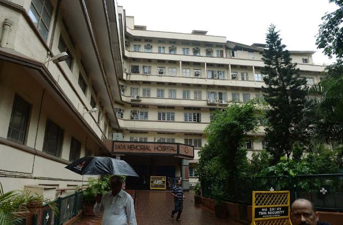
Inner Banner

Arvind Ingle group
Officer-in-Charge: Dr. Arvind Ingle

The Histology facility provides the following services to the Centre: (a) slides of unstained/ haematoxylin and eosin (H&E) stained histology sections of animal tissues including bone/ tumour samples, (b) logistic support for frozen sectioning of human/ animal tissues, and (c) blocks of multiple tissues by pecking method using a microarray machine. During 2017, the facility received 3471 tissue samples in fixative and 1231 human tissue paraffin blocks and, after processing, supplied 6869 stained and 17681 unstained slides to 20 research labs. In addition, 3397 tissues were processed for cryo-sectioning, and 1318 H&E stained and 1318 unstained slides were supplied to 12 research labs.

Dr. Sanjeev Waghmare
Officer-in-Charge: Dr. Sanjeev Waghmare
About Us

The Flow Cytometry (FCM) facility is a centralized facility used by scientists/ clinicians from ACTREC for a wide range of research applications including Immunophenotyping, Multicolor analysis, DNA content and cell cycle analysis, Apoptosis and Proliferation studies, Detection of mitochondrial membrane potential, Stem cell analysis, Detection of circulating tumor cells, Functional assays like Intracellular calcium influx, Oxidative burst analysis, Intracellular cytokine analysis, Cytometric bead array assay for the detection of cytokines, and 4-way live cell sorting.

The facility provides technical expertise in experiment design and data interpretation to researchers whenever required, and also provides training in data analysis.

The facility has four flow cytometers: FACSAria III, FACSAria I, Attune NxT and FACSCalibur. FACSAria III is equipped with 5 lasers (UV: 355 nm, Violet: 405 nm, Blue: 488 nm, Yellow green: 561 nm and Red: 633 nm) and can perform 18 color analysis. FACSAria-I is equipped with 3 lasers (Violet: 405 nm, Blue: 488 nm and Red: 633 nm) and can analyse up to 11-color. Both the Cell sorter can perform 4-way cell sorting as well as single cell sorting into 96-well plates using the Automated Cell Deposition System. Attune NxT is equipped with 4 lasers (Violet: 405 nm, Blue: 488 nm, Yellow green: 561 nm and Red: 633 nm) and can analyse up to14 color. FACSCalibur is equipped with one laser (Blue: 488 nm) and can perform 3-color analysis.

The software used for data analysis include FlowJo, FACSDiva, CellQuest Pro, Attune NxT, FCAP Array and Modfit.

The facility also offers its services to outside investigators on payment basis. Demonstrations and training were provided to staff and students on request.

Services

Flow cytometry facility is a centralised facility provides technical resources and professional expertise for multicolour flow cytometry analysis and cell sorting.

The following services has been provided by Facility:

  • Multicolor analysis can be done efficiently up to 18 colors by FACSAria III (BD Biosciences) and 14 colors by using Attune NxT (Life Technologies).
  • Cell sorting can be done by FACSAria I (up to 11 Colors and FACSAria III (up to 18 Colors). Both the cell sorters can sort 4 different populations at a time and also perform plate sorting for clonal analysis.
  • Software Flow Jo, FACSDiva, ModFit (Cell cycle analysis), Attune NxT, Cell Quest Pro, FCAP Array (Cytometric Bead Array Assay analysis) can be used for analysis purpose.
  • The technical support is extended to design the flow cytometry based experiments, to optimise the reagents (e.g. antibodies concentration), Florochrome selection and further assistance in data analysis.

For External User

The Flow cytometry facility at ACTREC is open to external user with certain conditions as follows:

  • Booking has to be done at least one week prior in advance and depending upon the availability of the slot, booking will be available to external user.
  • User should prepare the sample in their own Institute/ Lab and bring it in sterile condition.
  • It is recommended to pass the sample through strainer to avoid clogging of sample line.
  • The user should bear the cost of analysis of data separately.
  • The data should be collected immediately after acquisition. Users are requested to carry CD to copy the data.

Charge for Outside Users

Government Institutes/ University   - Rs. 600/ Tube (for acquisition)
Corporate                  - Rs. 2000/ Tube (for acquisition)

The user should contact of any query to Flow Cytometry facility on following:

Dr. Sanjeev Waghmare
Email: swaghmare@actrec.gov.in
      flowcytometry@actrec.gov.in
Contact: 022-27405000 Ext. 5316/5122

Protocol

DNA Cell Cycle using Propidium iodide

Reagents and Instruments

  • Cell lines or primary cell cultures
  • Phosphate buffered saline (0.01 M PBS pH 7.4)
  • 70% ethanol
  • Propidium iodide (PI)- Stock 400 μg/ml
  • RNase- Stock 1mg/ml
  • Triton X-100
  • Polystyrene round bottom 12 x 75 mm Falcon tubes (Flow Tubes)
  • 1 ml syringes with 26 G (Guage) needle
  • Flow cytometer- FACSCalibur, Attune Nxt
  • Softwares – CellQuest Pro, ModFit

Sample fixation

  1. Dispense 1 x 10⁶ cells per ml of PBS in 5ml polystyrene tubes.
  2. Wash the cells with 1 ml of PBS by centrifuging at 1000 rpm for 10 mins.
  3. Fix the cells by adding 70% chilled ethanol drop wise to the cell pellet while vortexing. This should ensure fixation of all cells and minimize clumping.
  4. Keep cells on ice for 1 hr or overnight at 4℃. (Fixed cells can be stored at 4℃ for 15-20 days)

Sample Staining

Procedure – I
  1. Take 1 x 10⁶ fixed cells in 5ml polystyrene tubes and centrifuge at 1000 rpm for 10 mins.
  2. Discard supernatant, wash the cells with 1 ml of PBS by centrifuging at 1000 rpm for 10 mins.
  3. Add 500 μl of PBS to the cell pellet, vortex gently, add 100 μl of Propidium Iodide and 100 μl of RNase. Cover the tubes with aluminum foil to prevent its exposure to light. Incubate at 37ºC for 30 mins.
  4. Pass the sample through 26 G needle to prepare single cell suspension.
  5. Acquire the sample on FACSCalibur / Attune NxT.
Procedure- II
  1. Take 1 x 10⁶ fixed cells in 5ml polystyrene tubes and centrifuge at 1000 rpm for 10 mins.
  2. Discard the supernatant and wash the cells with 1 ml of PBS by centrifuging at 1000 rpm for 10 mins
  3. Re-suspend the pellet in 500 μl of PBS containing 0.1% Triton X-100 and 100 μg/ml of RNaseA for permeabilization and RNase inactivation respectively. Incubate at 37ºC for 15 mins.
  4. After incubation, add 25 μg/ml of propidium iodide to the cell suspension, mix properly and further incubate at 37°C for 15 minutes.
  5. Pass the cell suspension through 26 G needle to avoid clumping of sample.
  6. Acquire the sample on FACSCalibur / Attune NxT.

Preparation of reagents

1. Phosphate buffer saline (0.01 M PBS pH 7.4)

NaCl8.0 g
KCl0.2 g
Na2HPO41.44 g
KH2PO40.24 g

Dissolve in 800 ml distilled water (dH2O)
Adjust the pH to 7.4 with HCl
Finally, make up the volume to 1 L with dH2O.
Store at 4°C.

2. 70 % ethanol

Ethanol70 ml
Distilled water (dH2O)30 ml

Store at 4°C.

3. Propidium iodide (PI): Stock – 400 μg /ml

Propidium iodide (Sigma)4 mg
PBS (0.01 M, pH 7.4)10 ml

Dissolve and Aliquots in 1.5 ml Eppendorf tubes
Store at -20°C.
(Precaution: Cover the 1.5 ml Eppendorf tubes with Aluminum foil as PI is light sensitive.

4. RNase: Stock – 1 mg/ml

RNase (Sigma)1 mg
PBS (0.01 M, pH 7.4)1 ml

Dissolve and aliquot in Eppendorf tubes.
Store at -20°C.

Point to remember:

  1. The quality of sample should be maintained for better result.
  2. Try to make single cell suspension before fixation of the sample to avoid clumping (to prevent the doublets).
  3. Handle the tubes with care and wear gloves to avoid contact with skin as PI is mutagenic.

Immunophenotyping

There are three general staining protocols based on type of antigen to be evaluate, nature of Fluorochrome and sample type.

  1. Direct Staining:

    This technique involves incubation of cells with fluorochrome conjugated antibody, which recognize and bind to various cell surface antigens. The antibodies are mostly monoclonal antibodies (MAb) and they may be added either single or in multiple combinations depending upon the analysis.

  2. Indirect staining:

    In indirect staining technique, the cells are incubated with an unconjugated primary antibody. The primary antibody recognizes and binds to cell surface antigens then the cells are subsequently incubated with a fluorochrome conjugated secondary antibody.

  3. Intracellular staining:

    This technique involves detection of intracellular antigens; it done by direct staining method using conjugated antibodies. Cells are initially subjected to a fixation step with paraformaldehyde in order to minimize leakage of proteins out of the cell, following permeabilization by mild detergents such as saponin or Tween 20. The permeabilization allows access of conjugated antibodies to proteins within the cell. The conjugated antibody penetrates and binds to its target within the cell. Intracellular staining technique is very useful in monitoring immune responses and tracking signalling molecules.

Reagents and Instruments

  • Cell line or primary cells
  • Phosphate buffered saline (PBS, 0.01M pH 7.4)
  • FACS Buffer I (for surface staining)
  • FACS Buffer II (for intracellular staining)
  • 1% Paraformaldehyde
  • Refrigerated centrifuge
  • Polystyrene round bottom 12 x 75 mm Falcon tubes
  • Micropipettes of capacity- 1 ml, 200 μl, 10 μl
  • Flow cytometer – FACSCalibur, Attune NxT or FACSAria
  • Software - CellQuest Pro (FACSCalibur), Attune NxT (Attune NxT), FACSDiva (FACSAria)
Antibodies

Direct staining:
1. Fluorochrome labelled antibody
2. Isotype matched fluorochrome labelled antibody
Indirect staining:
1. Unlabelled primary antibody
2. Fluorochrome labelled secondary antibody

Procedure

  1. Direct Staining
    1. Dispense 1 x 10⁶ Cells in 5ml polystyrene tubes.
    2. Add 1 ml of FACS buffer I and centrifuge the tubes at 1000 rpm for 10 mins at 4°C. Discard the supernatant. (washing)
    3. Resuspend the cells in 100 μl of FACS buffer I.
    4. Add appropriate amount of antibody (the amount of antibody can be decided as per manufacturer’s recommendation and titration)
    5. As negative control, incubate the cells with isotype matched antibody conjugated with same fluorochrome used for the test.
    6. Incubate the cells on ice at 4°C for 45 mins. in dark.
    7. Wash the cells 2 times and finally resuspend in 500 μl of FACS buffer I.
    8. Acquire the sample on flow cytometer as soon as possible.
    9. If the acquisition is not possible on the same day, fix the cells in 500 μl of 1% paraformaldehyde to preserve the cells for several days (this will stabilize the light scatter, prevent quenching and inactivate most biohazardous agents).
  2. Indirect Staining
    1. Dispense 1 x 10⁶ Cells in 5ml polystyrene tubes.
    2. Add 1 ml of FACS buffer I and centrifuge the tubes at 1000 rpm for 10 min at 4°C. Discard the supernatant (washing).
    3. Resuspend the cells in 100 μl of FACS buffer I.
    4. Add appropriate amount of unconjugated purified monoclonal antibody specific for antigen to be detected. (the amount of antibody can be change based on manufacturer’s recommendation and titration)
    5. Incubate on ice at 4 0 C for 45 mins.
    6. Wash the cells 2 times with FACS buffer I as described before to remove excess antibody.
    7. Dilute the fluorochrome- labelled secondary antibody as per manufacturer’s instruction in FACS buffer I.
    8. Incubate the cells with diluted secondary antibody (specific for primary antibodies) conjugated with fluorochrome on ice at 40 C for 45 min.
    9. As negative controls, incubate the cells with isotype matched controls conjugated with same fluorochrome used for the test.
    10. Wash the cells 2 times and finally resuspend in 500 μl of FACS buffer I.
    11. Acquire the sample on flow cytometer as soon as possible.
    12. If the acquisition is not possible on the same day, fix the cells in 500 μl of 1% paraformaldehyde to preserve the cells for several days (this will stabilize the light scatter, prevent quenching and inactivate most biohazardous agents).
  3. Intracellular staining
    1. Dispense cells in 5ml tubes.
    2. Add 1 ml of FACS buffer I and centrifuge the tubes at 1000 rpm for 10 mins at 40C. Discard the supernatant (washing).
    3. Add 500 μl of 1 % paraformaldehyde to fix the cells.
    4. Incubate on ice at 40C for 15 mins. with intermediate mixing of the tubes.
    5. Wash the cells with FACS buffer I by centrifuging tubes at 1000 rpm for 10 mins. at 40C twice.
    6. After second wash, Add 100 μl FACS buffer II to the cell pellet to permeabilize the cells.
    7. Gently vortex the tubes.
    8. Incubate at room temperature for 5 min.
    9. Add recommended amount of fluorochrome labelled antibody specific for antigen to be detected.
    10. As negative control, incubate the cells with isotype matched controls conjugated with same fluorochrome used for the test.
    11. Incubate on ice at 4°C for 45 mins. in dark.
    12. Wash the cells 2 times and resuspend in 500 μl of FACS buffer I.
    13. Acquire the sample on flow cytometer.

Preparation of reagents:

1. Phosphate buffer saline (0.01 M PBS pH 7.4)

NaCl8.0 g
KCl0.2 g
Na2HPO41.44 g
KH2PO40.24 g

Dissolve in 800 ml distilled water (dH2O)
Adjust the pH to 7.4 with HCl
Finally, make up the volume to 1 L with dH2O.
Store at 4°C.

2. FACS buffer I

PBS (0.01 M, pH 7.4)99 ml
Fetal Bovine Serum (Invitrogen)1.0 ml
Sodium Azide (NaN3)0.02 g

Store at 4°C.

3. FACS buffer II

PBS (0.01 M, pH 7.4)99 ml
Fetal Bovine Serum1.0 ml
Sodium Azide (NaN3)0.02 g

Store at 4°C.

4. 1 % paraformaldehyde

Paraformaldehyde1.0 g
PBS (0.01 M, pH 7.4)100 ml

Prepare solution in a glass bottle. Heat in microwave oven until the powder dissolves completely. Allow it to cool. Cover the bottle with Aluminium foil to prevent exposure to light. Store at 4°C.
(Precaution: Do not inhale the fumes)

Point to remember:

  1. Before starting the experiment, the titration of antibodies should be done according to manufacturer’s instruction for optimum concentration.
  2. The recommended controls should be run with every experiment for better analysis e.g. Iso control
  3. In case of multicolour analysis use compatible compensation controls.

Annexin V / Propidium iodide staining (For Apoptosis)

Reagents and Instruments:

  • Phosphate buffered saline (0.01 M PBS pH 7.4)
  • Annexin-V FITC
  • Propidium iodide (PI) – stock- 400 μg/ml
  • Annexin Binding buffer- 10X
  • Refrigerated centrifuge
  • Polystyrene round bottom 12 x 75 mm Falcon tubes
  • Micropipettes of capacity- 1 ml, 200 μl, 10 μl
  • Flow cytometer- FACSCalibur, Attune Nxt
  • Softwares – CellQuest Pro, Attune NxT, Flow Jo

Procedure:

  1. Wash the cells twice with 1X PBS and then resuspend in 1X Annexin Binding Buffer at a concentration of 1 X 10⁶ cells/ml.
  2. Transfer 100 μl of the cell suspension (1 X 10⁵ cells) in another polystyrene tube.
  3. Add 2-5 μl of Annexin V FITC.
  4. Add 40-50 μg of Propidium Iodide.
  5. Gently vortex the cells and incubate for 15 min at room temperature in the dark.
  6. Add 300 μl of 1X Annexin Binding Buffer to each tube. Analyze by flow cytometry within 1 hr.
The following controls are used to set up compensation
  1. Unstained cells.
  2. Cells stained with only Annexin V FITC
  3. Cells stained with only Propidium Iodide

Preparation of Reagents:

1. Phosphate buffer saline (0.01 M PBS pH 7.4)

NaCl8.0 g
KCl0.2 g
Na2HPO41.44 g
KH2PO40.24 g

Dissolve the following in 80ml MilliQ Water:

2. Annexin binding buffer 10X

HEPES (0.1 M)2.603 g
KCl0.2 g
cacl20.368 g
NaCl (1.4 M)8.182 g

Adjust the pH to 7.4. Make up the volume to 100 ml with MilliQ water. Store at 2-8°C

3. Propidium iodide (PI): Stock – 400 μμg /ml

Propidium iodide (Sigma)4 mg
PBS (0.01 M, pH 7.4)10 ml

Dissolve and aliquot in 1.5 ml capacity tubes.
Store at -20°C.
(Precaution: Cover the tube with aluminium foil as PI is photo-sensitive.)

Point to remember:

  1. Before starting the experiment, the titration of Annexin V should be done to decide optimal concentration.
  2. The pH of Annexin binding buffer should be maintained for better results.
  3. Handle with tubes with care and wear gloves to avoid contact with skin as PI is mutagenic.
User Guidelines

SOP for Flow Cytometry Facility User

New Flow Cytometry facility users are required to fill up the Project details form before starting the experiment. A hard copy of the same should be submitted with signatures of Principal Investigator/Officer-in-charge/ Scientific officers. The format will be available at the Facility.

Booking for Acquisition and Analysis of samples

  1. Booking has to be done in Booking register that is available in the Facility.
  2. Booking can be done one-week prior to the date of sample acquisition.
  3. Booking slots are available for both acquisition and sorting. In a given day, only one booking slot of 2 hrs is allowed. Additional time will be entertained based on the nature of the experiments and number samples depending on the availability of the slots.
  4. Cancellation of booking if any should be informed at least one-day prior of date of acquisition.
  5. If any queries, please contact Facility staff.

Sample Preparation for Flow Cytometry

  1. Try to prepare single cells suspension and avoid clumping. Users are Recommended to pass the cells through filters or strainers or insulin syringe.
  2. Sample concentration should be maintained for better results. (e.g. 1 X10⁶ Cells / 300-500 μl for acquisition depending upon the cell type)
  3. For sorting carry sufficient number of collection tubes with appropriate quantity of media or buffer.
  4. User can use 15 ml tubes, 5 ml FALCON tubes, Eppendorf 1.5 /2 ml, and 96 well plates for collection of cells.

General Guidelines

  • Suitable Controls should be carry at the time of acquisition and if any queries please contact Facility Staff.
  • Data can be collected in FCS format in the CD immediately after the sample acquisition.
  • Users can use softwares Flowjo, FACS DIVA, Attune NxT, Modfit LT 5.0, Cells Quest Pro, for analysis with prior booking.
Contact Us

Dr. Sanjeev K Waghmare
Principal Investigator
Scientific Officer’ F’
Officer In-Charge
Email: swaghmare[at]actrec[dot]gov[dot]in
Phone:022-27405122

Flow Cytometry Facility, ACTREC
Email: flowcytometry[at]actrec[dot]gov[dot]in
Phone: 022-27405000 Ext. 5316

Dr.Sorab N. Dalal
Officer-in-Charge: Dr.Sorab N. Dalal

The Electron Microscope Facility at ACTREC is equipped with JEM 1400 PLUS, a Transmission electron microscope (JEOL, Japan) functioning at 120kV with magnification up to 12,00,000 X and 0.2nm resolution. The facility provides services for research and analysis of biological samples, nanoparticles and other applications.

Current Applications
  • The facility carries out sample preparation for routine electron microscopy, including resin block making (solid tissues, monolayer cell cultures, and cell suspensions) followed by ultrathin sectioning using Leica UC-7 ultramicrotome, contrasting, and acquisition.
  • The facility also carries out negative staining for small particles (<100nm) like bacteriophages, exosomes, nanoparticles, proteins and DNA.
  • The facility provides acquisition of immunogold labelled macromolecules to detect specific localization.
  • The facility provides elemental detection by Bruker EDS system

The major areas of electron microscopy analysis are,

  • Ultrastructure study of cell and tissue,
  • Studying the details at organelle level like mitochondrial changes, ER - mitochondrial interactions, mitochondrial cristae, cell membrane, nuclear architecture, golgi complexes etc.,
  • To check events of autophagy, apoptosis or necroptosis,
  • Characterization of exosomes, bacteriophages and nanoparticles,
  • Uptake and effect of nanoparticles / drugs / radiation on cells or tissue,
  • EDS analysis of nanoparticles
Sample Submission

Please write to the facility regarding your requirement for electron microscopy. The facility will provide the details as per the analysis requirement. The users are requested to discuss over email regarding their study and sample submission. The samples need to be submitted along with a duly filled requisition form. The requisition form will be provided by the facility on request.

Charges
ACTREC & DAEOther than ACTREC institutionsCorporate
Routine EM1000500010000
Routine EM + Immunogold labeling1500600012000
Negative staining80040008000

18% GST to be included in addition to the following charges.

Mode of Payment

The charges should be paid against the quotation or invoice to the following account. The epayment details will be provided by the facility on request. The transaction details should be sent by email to the facility. The acquisition will be carried out after the receipt of the payment is confirmed.

Contact

Office Incharge:Dr Sorab N. Dalal
SO ‘H’
ACTREC, TMC

Operator-in-charge: Siddhi A. Redkar
SA ‘E’
Electron Microscope Facility
(Khanolkar Shodhika - 68)
ACTREC, TMC
Plot No.1 & 2, Sector 22
Kharghar, Navi Mumbai - 410 210.
022-27405000 / 022-68735000
Extension : 5545
Email : tem.facility@actrec.gov.in

Media Gallery
CellCell
Cell to cell ContactCell
CellCell
ExosomesMitochondria
CellCell
NanoparticlesGolgi Complex

Dr. Pradnya Kowtal
Officer-in-Charge: Dr. Pradnya Kowtal

The DNA sequencing facility has two automated DNA sequencers: an eight capillary Genetic Analyzer 3500 and a 48 capillary Genetic Analyzer 3730 from Applied Biosystems/ Thermofisher, both of which are used for DNA sequencing and fragment analysis. The average turnaround time to give out data is one working day after receiving samples. During the year 2017, the facility carried out a total of 25,459 reactions of which 13,718 were for sequencing and 11,741 were for fragment analysis. The facility also provided demonstration of Sanger sequencing to students and other visitors.

Dr. Dibyendu Bhattacharyya
Officer-in-Charge: Dr. Dibyendu Bhattacharyya

The ACTREC Digital Imaging facility (ADIF) is a state of the art imaging facility housing several advanced imaging platforms. At present, the facility boasts of the following high end instrumentation:

  • LSM510 confocal microscope
  • Multiphoton confocal LSM780 microscope
  • 3i Mariana spinning disk confocal microscope
  • Leica SP8 confocal microscope with STED super resolution system
  • Leica DMI600B microscope - from Bhattacharyya lab
  • Axio Imager Z1
  • Axiovert 200M

The facility provides microscopic acquisition and analysis services for wide-field and confocal platforms listed above, to the ACTREC faculty and students, and also to outsider users.

Mr. Uday Dandekar
Officer-in-Charge: Mr. Uday Dandekar

Over the past 40 years, the Institute has maintained a Common Instrument Room as a facility housing vital scientific equipment that are routinely required by the Centre’s staff and students, to optimize their utilization and make them available round the clock on all days of the week - including holidays. The facility also provides technical guidance and support to various research laboratories in the procurement and maintenance of their capital equipment.

Technically qualified staff members attached to this facility handle routine maintenance of all the equipment and render help to the end users, thus ensuring proper use of the equipment.

Requisite spares for centrifuges, low temperature freezers, CO2 incubators, etc. and consumables like centrifuge tubes, thermal paper rolls, etc. are procured on a regular basis and kept in stock in the facility to reduce downtime of the equipment. In all, 94 equipments are currently housed in this facility. During 2017, a multimode microplate reader was procured and installed in the CIR.

Dr. Abhijit De
Officer-in-Charge: Dr. Abhijit De

Established in 2013, this facility provides preclinical in vivo molecular imaging services to the ACTREC researchers. Molecular imaging, which provides real-time visualization and quantitative measurement of cellular processes at the molecular/ genetic level, adds value for translating the basic research findings to the clinic. Several in house research laboratories are using this facility for cancer therapeutic applications.

The facility has also supported researchers from neighboring institutions like IIT-Bombay. Instrumentation in this facility procured through various extramural grants include an IVIS Lumina II and IVIS Spectrum imaging system (both from Perkin Elmer, USA), a data server, two computer terminals to store and analyse imaging data, and additional gas anesthesia systems for optimal operation and use of this facility.

The installed systems offer fast scanning of multiple mice, rats or other small animals emitting photon signal from various sources such as bioluminescence, near-infrared fluorescence and Cerenkov luminescence. Salient features of these systems include high-performance, user-friendly acquisition and fully software-controlled image capture; data back-up storage server linked through ACTREC LAN as well as onsite and remote image processing units.

The systems are integrated with a heated stage and accessories for isoflurane based gas anesthesia needed for the non-invasive scanning procedure; they provide the ability to scan and quantify fluorescent, bioluminescent as well as Cerenkov signals (around 400–900 nm) from tissue culture plates, tubes or mice.

There is an integrated fluorescence system for switching between fluorescent and bioluminescent spectral imaging. The excitation/ emission filters accommodate majority fluorescent dyes or fluorescent proteins in the green to far-red spectral range. Spectral imaging options to obtain data from a sequence of images at different wavelengths in the visible range for determining the location of bioluminescent reporter. The filters can distinguish reporters with different wavelengths coming out from the same animal. Another important feature is the laser scanner for 3D surface topography to develop single-view diffuse tomographic reconstructions (DLIT and FLIT mode).

Dr. Sanjay Gupta
Officer-in-Charge: Dr. Sanjay Gupta

The Common Facilities (CF) operates and maintains support services like X-ray developing machine, ultra-pure water purification system, radioactive handling room for 32P and 125I, bacterial culture hoods, ice making machines and cold room facility to different research groups. In addition, maintenance of all the autoclaves and ovens belonging to different research groups is handled by the CF technicians.

All the major equipment under common facility is covered under annual maintenance contract in order to provide safe, sustainable, efficient and reliable facilities.

Dr. Poonam Gera
Officer-in-Charge: Dr. Poonam Gera

The ACTREC Biorepository is the custodian of stored biological samples that can be shared, under a tightly regulated and strictly monitored mechanism, with researchers having approved projects that aim to study the biology of cancer, find biomarkers for a more refined molecular classification, or for targeted therapy. In all, tissue samples from 630 cases were accrued in the Biorepository during 2017. As always the majority were head and neck tumors, followed by breast tumors. Other tumor types included neurological, gastrointestinal, genitourinary, gynecological, etc.

This year, the collection of bio specimens was extended to the Breast OPD wherein core biopsies are now being cryo preserved for future research; this is in addition to collection from the OTs, Frozen room and Surgical Pathology. Cryopreserved tissue samples (470) were provided to eight Principal Investigators with approved projects under various protocols at the Tata Memorial Centre. In the International Cancer Genome Consortium (ICGC) project, gingivo buccal mucosa tumour and blood samples were collected from 30 patients accrued this year, and their extracted genomic DNA samples were sent to NIBMG, Kalyani for whole genome scan and sequence capture-based flow cell sequencing.

The Biorepository itself initiated a project on quality monitoring of in-house cryo preserved tissues. The OIC contributed her Pathology expertise involving the evaluation of Hematoxylin & Eosin as well as immunohistochemistry slides to eight ACTREC projects.

Dr. Ashok Varma
Officer-in-Charge: Dr. Ashok Varma

The Bioinformatics facility of ACTREC provides infrastructural and technical support to scientists, clinicians and research scholars of the Centre to fulfil the bioinformatics requirements of their on-going research projects. Scientists of the Centre also use the facility’s infrastructure to explore microarray, next generation sequence data analysis, database development, molecular modelling and data mining for their on-going projects.

This facility receives strong funding support from DBT and is established as a BTIS-net centre of this region. It is well equipped with one nVIDIA Tesla GPU workstation, 5 workstations, 1 webserver and seven PCs. The facility also focuses on database development such as Histome: the human infobase; this database is presently being updated. In the domain of gene expression studies, projects related to TGCA database mining and analysis are under progress. In silico molecular modelling, dynamics and protein-protein interactions have been performed using BARC’s supercomputing facility.

The facility hosted a national 28th BTISnet Coordinators Meeting on 3rd and 4th February 2017. The facility also organized its annual 2-day Workshop on ‘Basics of Bioinformatics’ targeting college teachers and research scholars of institutions in the Mumbai and neighboring areas on 2nd and 3rd March 2017. The facility staff also provided training to six trainees during the year, three for Bachelor’s/ Master’s dissertation and three for experience.

Back to Top